


 Foundational characteristics of cancer include proliferation, angiogenesis, migration, evasion of apoptosis, and cellular immortality. Find key markers for these cellular processes and antibodies to detect them.
 The SUMOplot™ Analysis Program predicts and scores sumoylation sites in your protein. SUMOylation is a post-translational modification involved in various cellular processes, such as nuclear-cytosolic transport, transcriptional regulation, apoptosis, protein stability, response to stress, and progression through the cell cycle.
 The Autophagy Receptor Motif Plotter predicts and scores autophagy receptor binding sites in your protein. Identifying proteins connected to this pathway is critical to understanding the role of autophagy in physiological as well as pathological processes such as development, differentiation, neurodegenerative diseases, stress, infection, and cancer.
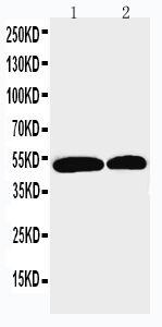
Anti-NOXA1 Antibody
- SPECIFICATION
- CITATIONS
- PROTOCOLS
- BACKGROUND

Application
| WB |
|---|---|
| Primary Accession | Q86UR1 |
| Host | Rabbit |
| Reactivity | Human |
| Clonality | Polyclonal |
| Format | Lyophilized |
| Description | Rabbit IgG polyclonal antibody for NADPH oxidase activator 1(NOXA1) detection. Tested with WB in Human. |
| Reconstitution | Add 0.2ml of distilled water will yield a concentration of 500ug/ml. |
| Gene ID | 10811 |
|---|---|
| Other Names | NADPH oxidase activator 1, NOX activator 1, Antigen NY-CO-31, NCF2-like protein, P67phox-like factor, p51-nox, NOXA1, P51NOX |
| Calculated MW | 50933 MW KDa |
| Application Details | Western blot, 0.1-0.5 µg/ml, Human |
| Subcellular Localization | Cytoplasm. Cell membrane. Translocation to membranes depends on NOXO1 or NCF1 and maybe RAC1. |
| Tissue Specificity | Widely expressed. Detected in pancreas, liver, kidney, spleen, prostate, small intestine and colon. . |
| Protein Name | NADPH oxidase activator 1 |
| Contents | Each vial contains 5mg BSA, 0.9mg NaCl, 0.2mg Na2HPO4, 0.05mg Thimerosal, 0.05mg NaN3. |
| Immunogen | A synthetic peptide corresponding to a sequence in the middle region of human NOXA1(176-195aa RQVPRGEVFRPHRWHLKHLE). |
| Purification | Immunogen affinity purified. |
| Cross Reactivity | No cross reactivity with other proteins |
| Storage | At -20˚C for one year. After r˚Constitution, at 4˚C for one month. It˚Can also be aliquotted and stored frozen at -20˚C for a longer time.Avoid repeated freezing and thawing. |
| Sequence Similarities | Belongs to the NCF2/NOXA1 family. |
| Name | NOXA1 |
|---|---|
| Synonyms | P51NOX |
| Function | Functions as an activator of NOX1, a superoxide-producing NADPH oxidase. Functions in the production of reactive oxygen species (ROS) which participate in a variety of biological processes including host defense, hormone biosynthesis, oxygen sensing and signal transduction. May also activate CYBB/gp91phox and NOX3. |
| Cellular Location | Cytoplasm. Cell membrane. Note=Translocation to membranes depends on NOXO1 or NCF1 and maybe RAC1 |
| Tissue Location | Widely expressed. Detected in pancreas, liver, kidney, spleen, prostate, small intestine and colon |

Thousands of laboratories across the world have published research that depended on the performance of antibodies from Abcepta to advance their research. Check out links to articles that cite our products in major peer-reviewed journals, organized by research category.
info@abcepta.com, and receive a free "I Love Antibodies" mug.

Provided below are standard protocols that you may find useful for product applications.
Background
NOXA1(NADPH oxidase activator 1), also called NOX ACTIVATOR 1 or p51-NOX, is an enzyme that in humans is encoded by the NOXA1 gene. Hartz(2007) mapped the NOXA1 gene to chromosome 9q34.3 based on an alignment of the NOXA1 sequence with the genomic sequence(build 36.1). Banfi et al.(2003) mapped the mouse Noxa1 gene to chromosome 2. Using yeast 2-hybrid assays, Takeya et al.(2003) showed that human p51-NOX interacted with constitutively active forms of RAC1 and RAC2. In vitro binding assays revealed that p51-NOX bound GTP-bound RAC1, but not GDP-bound RAC1. p51-NOX also bound p47-PHOX(NCF1) and p41-NOX(NOXO1), and trp436 within the SH3 domain of p51-NOX was required for these interactions. Human cell lines or COS-7 cells cotransfected with p51-NOX and p41-NOX and either gp91-PHOX(CYBB) or NOX1 produced superoxide. Cells individually transfected with NOX1, p41-NOX, or p51-NOX and cells transfected with only p41-NOX and p51-NOX showed no superoxide production.
If you have used an Abcepta product and would like to share how it has performed, please click on the "Submit Review" button and provide the requested information. Our staff will examine and post your review and contact you if needed.
If you have any additional inquiries please email technical services at tech@abcepta.com.
Ordering Information
Other Products
Shipping Information
