


 Foundational characteristics of cancer include proliferation, angiogenesis, migration, evasion of apoptosis, and cellular immortality. Find key markers for these cellular processes and antibodies to detect them.
 The SUMOplot™ Analysis Program predicts and scores sumoylation sites in your protein. SUMOylation is a post-translational modification involved in various cellular processes, such as nuclear-cytosolic transport, transcriptional regulation, apoptosis, protein stability, response to stress, and progression through the cell cycle.
 The Autophagy Receptor Motif Plotter predicts and scores autophagy receptor binding sites in your protein. Identifying proteins connected to this pathway is critical to understanding the role of autophagy in physiological as well as pathological processes such as development, differentiation, neurodegenerative diseases, stress, infection, and cancer.
Anti-PON1 Antibody
- SPECIFICATION
- CITATIONS
- PROTOCOLS
- BACKGROUND

Application
| WB |
|---|---|
| Primary Accession | P27169 |
| Host | Rabbit |
| Reactivity | Human, Mouse, Rat |
| Clonality | Polyclonal |
| Format | Lyophilized |
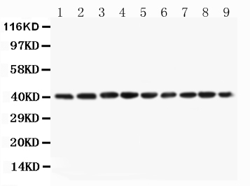
| Description | Rabbit IgG polyclonal antibody for Serum paraoxonase/arylesterase 1(PON1) detection. Tested with WB in Human;Mouse;Rat. |
| Reconstitution | Add 0.2ml of distilled water will yield a concentration of 500ug/ml. |
| Gene ID | 5444 |
|---|---|
| Other Names | Serum paraoxonase/arylesterase 1, PON 1, 3.1.1.2, 3.1.1.81, 3.1.8.1, Aromatic esterase 1, A-esterase 1, K-45, Serum aryldialkylphosphatase 1, PON1, PON |
| Calculated MW | 39731 MW KDa |
| Application Details | Western blot, 0.1-0.5 µg/ml, Human, Mouse, Rat |
| Subcellular Localization | Secreted, extracellular space. |
| Tissue Specificity | Plasma, associated with HDL (at protein level). Expressed in liver, but not in heart, brain, placenta, lung, skeletal muscle, kidney or pancreas. . |
| Protein Name | Serum paraoxonase/arylesterase 1 |
| Contents | Each vial contains 5mg BSA, 0.9mg NaCl, 0.2mg Na2HPO4, 0.05mg Thimerosal, 0.05mg NaN3. |
| Immunogen | A synthetic peptide corresponding to a sequence in the middle region of human PON1(186-201aa FLDPYLQSWEMYLGLA), different from the related mouse and rat sequences by three amino acids. |
| Purification | Immunogen affinity purified. |
| Cross Reactivity | No cross reactivity with other proteins |
| Storage | At -20˚C for one year. After r˚Constitution, at 4˚C for one month. It˚Can also be aliquotted and stored frozen at -20˚C for a longer time.Avoid repeated freezing and thawing. |
| Sequence Similarities | Belongs to the paraoxonase family. |
| Name | PON1 |
|---|---|
| Synonyms | PON |
| Function | Hydrolyzes the toxic metabolites of a variety of organophosphorus insecticides. Capable of hydrolyzing a broad spectrum of organophosphate substrates and lactones, and a number of aromatic carboxylic acid esters. Mediates an enzymatic protection of low density lipoproteins against oxidative modification and the consequent series of events leading to atheroma formation. |
| Cellular Location | Secreted, extracellular space. |
| Tissue Location | Plasma, associated with HDL (at protein level). Expressed in liver, but not in heart, brain, placenta, lung, skeletal muscle, kidney or pancreas. |

Thousands of laboratories across the world have published research that depended on the performance of antibodies from Abcepta to advance their research. Check out links to articles that cite our products in major peer-reviewed journals, organized by research category.
info@abcepta.com, and receive a free "I Love Antibodies" mug.

Provided below are standard protocols that you may find useful for product applications.
Background
Serum paraoxonase/arylesterase 1(PON1), also known as aromatic esterase 1, is an enzyme that in humans is encoded by the PON1 gene. It is mapped to 7q21.3. This gene has esterase and more specifically paraoxonase activity. PON1 is responsible for hydrolysing organophosphate pesticides and nerve gasses. Polymorphisms in the PON1 gene significantly affect the catalytic ability of the enzyme. PON1(paraoxonase 1) is also a major anti-atherosclerotic component of high-density lipoprotein(HDL). The PON1 gene is activated by PPAR-gamma, which increases synthesis and release of paraoxonase 1 enzyme from the liver, reducing atherosclerosis.
If you have used an Abcepta product and would like to share how it has performed, please click on the "Submit Review" button and provide the requested information. Our staff will examine and post your review and contact you if needed.
If you have any additional inquiries please email technical services at tech@abcepta.com.
Ordering Information
Other Products
Shipping Information
