


 Foundational characteristics of cancer include proliferation, angiogenesis, migration, evasion of apoptosis, and cellular immortality. Find key markers for these cellular processes and antibodies to detect them.
 The SUMOplot™ Analysis Program predicts and scores sumoylation sites in your protein. SUMOylation is a post-translational modification involved in various cellular processes, such as nuclear-cytosolic transport, transcriptional regulation, apoptosis, protein stability, response to stress, and progression through the cell cycle.
 The Autophagy Receptor Motif Plotter predicts and scores autophagy receptor binding sites in your protein. Identifying proteins connected to this pathway is critical to understanding the role of autophagy in physiological as well as pathological processes such as development, differentiation, neurodegenerative diseases, stress, infection, and cancer.
Anti-Cathepsin D Picoband Antibody
- SPECIFICATION
- CITATIONS
- PROTOCOLS
- BACKGROUND

Application
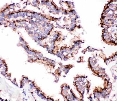
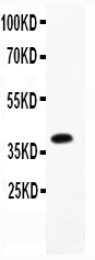
| WB, IHC-P |
|---|---|
| Primary Accession | P07339 |
| Host | Rabbit |
| Reactivity | Human |
| Clonality | Polyclonal |
| Format | Lyophilized |
| Description | Rabbit IgG polyclonal antibody for Cathepsin D(CTSD) detection. Tested with WB, IHC-P in Human. |
| Reconstitution | Add 0.2ml of distilled water will yield a concentration of 500ug/ml. |
| Gene ID | 1509 |
|---|---|
| Other Names | Cathepsin D, 3.4.23.5, Cathepsin D light chain, Cathepsin D heavy chain, CTSD, CPSD |
| Calculated MW | 44552 MW KDa |
| Application Details | Immunohistochemistry(Paraffin-embedded Section), 0.5-1 µg/ml, Human, By Heat Western blot, 0.1-0.5 µg/ml, Human |
| Subcellular Localization | Lysosome. Melanosome. Secreted, extracellular space. Identified by mass spectrometry in melanosome fractions from stage I to stage IV. In aortic samples, detected as an extracellular protein loosely bound to the matrix (PubMed:20551380). . |
| Tissue Specificity | Expressed in the aorta extrcellular space (at protein level). . |
| Protein Name | Cathepsin D |
| Contents | Each vial contains 5mg BSA, 0.9mg NaCl, 0.2mg Na2HPO4, 0.05mg NaN3. |
| Immunogen | E.coli-derived human Cathepsin D recombinant protein (Position: G65-L412). Human Cathepsin D shares 85% amino acid (aa) sequence identity with both mouse and rat Cathepsin D. |
| Purification | Immunogen affinity purified. |
| Cross Reactivity | No cross reactivity with other proteins |
| Storage | At -20˚C for one year. After r˚Constitution, at 4˚C for one month. It˚Can also be aliquotted and stored frozen at -20˚C for a longer time.Avoid repeated freezing and thawing. |
| Sequence Similarities | Belongs to the peptidase A1 family. |
| Name | CTSD |
|---|---|
| Synonyms | CPSD |
| Function | Acid protease active in intracellular protein breakdown. Plays a role in APP processing following cleavage and activation by ADAM30 which leads to APP degradation (PubMed:27333034). Involved in the pathogenesis of several diseases such as breast cancer and possibly Alzheimer disease. |
| Cellular Location | Lysosome. Melanosome. Secreted, extracellular space. Note=Identified by mass spectrometry in melanosome fractions from stage I to stage IV. In aortic samples, detected as an extracellular protein loosely bound to the matrix (PubMed:20551380) |
| Tissue Location | Expressed in the aorta extracellular space (at protein level) (PubMed:20551380). Expressed in liver (at protein level) (PubMed:1426530). |

Thousands of laboratories across the world have published research that depended on the performance of antibodies from Abcepta to advance their research. Check out links to articles that cite our products in major peer-reviewed journals, organized by research category.
info@abcepta.com, and receive a free "I Love Antibodies" mug.

Provided below are standard protocols that you may find useful for product applications.
Background
Cathepsin D is a protein that in humans is encoded by the CTSD gene. This proteinase is a member of the peptidase C1 family, having a specificity similar to but narrower than that of pepsin A. It is mapped to 11p15.5. The cDNA encodes a 412-amino acid protein with 20 and 44 amino acids in a pre- and prosegment, respectively. Cathepsin D is one of the lysosomal proteinases. It is ubiquitously expressed and is involved in proteolytic degradation, cell invasion, and apoptosis. Mutations in this gene are involved in the pathogenesis of several diseases, including breast cancer and possibly Alzheimer disease and it has been considered as a breast cancer tumor marker.
If you have used an Abcepta product and would like to share how it has performed, please click on the "Submit Review" button and provide the requested information. Our staff will examine and post your review and contact you if needed.
If you have any additional inquiries please email technical services at tech@abcepta.com.
Ordering Information
Other Products
Shipping Information
