


 Foundational characteristics of cancer include proliferation, angiogenesis, migration, evasion of apoptosis, and cellular immortality. Find key markers for these cellular processes and antibodies to detect them.
 The SUMOplot™ Analysis Program predicts and scores sumoylation sites in your protein. SUMOylation is a post-translational modification involved in various cellular processes, such as nuclear-cytosolic transport, transcriptional regulation, apoptosis, protein stability, response to stress, and progression through the cell cycle.
 The Autophagy Receptor Motif Plotter predicts and scores autophagy receptor binding sites in your protein. Identifying proteins connected to this pathway is critical to understanding the role of autophagy in physiological as well as pathological processes such as development, differentiation, neurodegenerative diseases, stress, infection, and cancer.
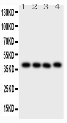
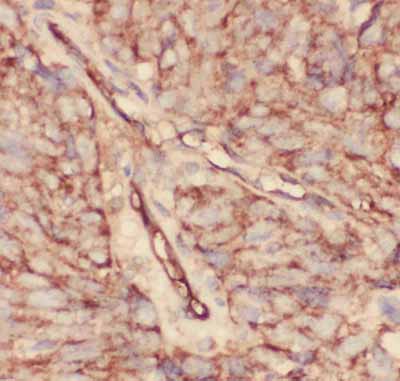
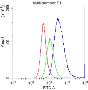
Anti-CD34 Picoband Antibody
- SPECIFICATION
- CITATIONS
- PROTOCOLS
- BACKGROUND

Application
| WB, IHC-P, IHC-F, FC, ICC |
|---|---|
| Primary Accession | P28906 |
| Host | Rabbit |
| Reactivity | Human, Mouse, Rat |
| Clonality | Polyclonal |
| Format | Lyophilized |
| Description | Rabbit IgG polyclonal antibody for Hematopoietic progenitor cell antigen CD34(CD34) detection. Tested with WB, IHC-P, IHC-F, ICC, FCM in Human;Mouse;Rat. |
| Reconstitution | Add 0.2ml of distilled water will yield a concentration of 500ug/ml. |
| Gene ID | 947 |
|---|---|
| Other Names | Hematopoietic progenitor cell antigen CD34, CD34, CD34 |
| Calculated MW | 40716 MW KDa |
| Application Details | Immunohistochemistry(Paraffin-embedded Section), 0.5-1 µg/ml, By Heat Immunohistochemistry(Frozen Section), 0.5-1 µg/ml Immunocytochemistry, 0.5-1 µg/ml Western blot, 0.1-0.5 µg/ml Flow Cytometry, 1-3μg/1x106cells |
| Subcellular Localization | Membrane; Single-pass type I membrane protein. |
| Tissue Specificity | Selectively expressed on hematopoietic progenitor cells and the small vessel endothelium of a variety of tissues. |
| Protein Name | Hematopoietic progenitor cell antigen CD34 |
| Contents | Each vial contains 5mg BSA, 0.9mg NaCl, 0.2mg Na2HPO4, 0.05mg NaN3. |
| Immunogen | E.coli-derived human CD34 recombinant protein (Position: T151-L385). Human CD34 shares 79% amino acid (aa) sequence identity with mouse CD34. |
| Purification | Immunogen affinity purified. |
| Cross Reactivity | No cross reactivity with other proteins |
| Storage | At -20˚C for one year. After r˚Constitution, at 4˚C for one month. It˚Can also be aliquotted and stored frozen at -20˚C for a longer time.Avoid repeated freezing and thawing. |
| Sequence Similarities | Belongs to the CD34 family. |
| Name | CD34 |
|---|---|
| Function | Possible adhesion molecule with a role in early hematopoiesis by mediating the attachment of stem cells to the bone marrow extracellular matrix or directly to stromal cells. Could act as a scaffold for the attachment of lineage specific glycans, allowing stem cells to bind to lectins expressed by stromal cells or other marrow components. Presents carbohydrate ligands to selectins. |
| Cellular Location | Membrane; Single-pass type I membrane protein. |
| Tissue Location | Selectively expressed on hematopoietic progenitor cells and the small vessel endothelium of a variety of tissues |

Thousands of laboratories across the world have published research that depended on the performance of antibodies from Abcepta to advance their research. Check out links to articles that cite our products in major peer-reviewed journals, organized by research category.
info@abcepta.com, and receive a free "I Love Antibodies" mug.

Provided below are standard protocols that you may find useful for product applications.
Background
CD34 is a monomeric cell surface antigen with a molecular mass of approximately 110 KD.CD34 is expressed in humans in hematopoietic stem cells, vascular endothelium, and blasts from 30% of patients with acute myeloid and lymphocytic leukemia. The human CD34 gene spans 26 kb and has 8 exons, a structure quite similar to that of the murine gene. By Southern blot analysis of DNA from a panel of human x mouse somatic cell hybrids using a CD34 cDNA probe demonstrate that the gene for CD34 is located on human chromosome 1 in the 1q12----qter region. CD34 plays an important role in the formation of progenitor cells during both embryonic and adult hematopoiesis.
If you have used an Abcepta product and would like to share how it has performed, please click on the "Submit Review" button and provide the requested information. Our staff will examine and post your review and contact you if needed.
If you have any additional inquiries please email technical services at tech@abcepta.com.
Ordering Information
Other Products
Shipping Information
