


 Foundational characteristics of cancer include proliferation, angiogenesis, migration, evasion of apoptosis, and cellular immortality. Find key markers for these cellular processes and antibodies to detect them.
 The SUMOplot™ Analysis Program predicts and scores sumoylation sites in your protein. SUMOylation is a post-translational modification involved in various cellular processes, such as nuclear-cytosolic transport, transcriptional regulation, apoptosis, protein stability, response to stress, and progression through the cell cycle.
 The Autophagy Receptor Motif Plotter predicts and scores autophagy receptor binding sites in your protein. Identifying proteins connected to this pathway is critical to understanding the role of autophagy in physiological as well as pathological processes such as development, differentiation, neurodegenerative diseases, stress, infection, and cancer.
Anti-CMA1 Picoband Antibody
- SPECIFICATION
- CITATIONS
- PROTOCOLS
- BACKGROUND

Application
| WB, IHC-P |
|---|---|
| Primary Accession | P23946 |
| Host | Rabbit |
| Reactivity | Human |
| Clonality | Polyclonal |
| Format | Lyophilized |
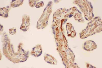
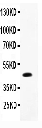
| Description | Rabbit IgG polyclonal antibody for Chymase(CMA1) detection. Tested with WB, IHC-P in Human. |
| Reconstitution | Add 0.2ml of distilled water will yield a concentration of 500ug/ml. |
| Gene ID | 1215 |
|---|---|
| Other Names | Chymase, 3.4.21.39, Alpha-chymase, Mast cell protease I, CMA1, CYH, CYM |
| Calculated MW | 27325 MW KDa |
| Application Details | Immunohistochemistry(Paraffin-embedded Section), 0.5-1 µg/ml, Human, By Heat Western blot, 0.1-0.5 µg/ml, Human |
| Subcellular Localization | Secreted. Cytoplasmic granule. Mast cell granules. |
| Tissue Specificity | Mast cells in lung, heart, skin and placenta. Expressed in both normal skin and in urticaria pigmentosa lesions. . |
| Protein Name | Chymase |
| Contents | Each vial contains 5mg BSA, 0.9mg NaCl, 0.2mg Na2HPO4, 0.05mg NaN3. |
| Immunogen | E.coli-derived human CMA1 recombinant protein (Position: I22-N247). Human CMA1 shares 75% and 74% amino acid (aa) sequences identity with mouse and rat CMA1, respectively. |
| Purification | Immunogen affinity purified. |
| Cross Reactivity | No cross reactivity with other proteins |
| Storage | At -20˚C for one year. After r˚Constitution, at 4˚C for one month. It˚Can also be aliquotted and stored frozen at -20˚C for a longer time.Avoid repeated freezing and thawing. |
| Sequence Similarities | Belongs to the peptidase S1 family. Granzyme subfamily. |
| Name | CMA1 |
|---|---|
| Synonyms | CYH, CYM |
| Function | Major secreted protease of mast cells with suspected roles in vasoactive peptide generation, extracellular matrix degradation, and regulation of gland secretion. |
| Cellular Location | Secreted. Cytoplasmic granule. Note=Mast cell granules |
| Tissue Location | Mast cells in lung, heart, skin and placenta. Expressed in both normal skin and in urticaria pigmentosa lesions |

Thousands of laboratories across the world have published research that depended on the performance of antibodies from Abcepta to advance their research. Check out links to articles that cite our products in major peer-reviewed journals, organized by research category.
info@abcepta.com, and receive a free "I Love Antibodies" mug.

Provided below are standard protocols that you may find useful for product applications.
Background
Chymase is a major secreted protease of mast cells with suspected roles in vasoactive peptide generation, extracellular matrix degradation, and regulation of gland secretion. This gene product is a chymotryptic serine proteinase that belongs to the peptidase family S1. Chymase is mapped to 14q12. It is expressed in mast cells and thought to function in the degradation of the extracellular matrix, the regulation of submucosal gland secretion, and the generation of vasoactive peptides. In the heart and blood vessels, this protein, rather than angiotensin converting enzyme, is largely responsible for converting angiotensin I to the vasoactive peptide angiotensin II.
If you have used an Abcepta product and would like to share how it has performed, please click on the "Submit Review" button and provide the requested information. Our staff will examine and post your review and contact you if needed.
If you have any additional inquiries please email technical services at tech@abcepta.com.
Ordering Information
Other Products
Shipping Information
