


 Foundational characteristics of cancer include proliferation, angiogenesis, migration, evasion of apoptosis, and cellular immortality. Find key markers for these cellular processes and antibodies to detect them.
 The SUMOplot™ Analysis Program predicts and scores sumoylation sites in your protein. SUMOylation is a post-translational modification involved in various cellular processes, such as nuclear-cytosolic transport, transcriptional regulation, apoptosis, protein stability, response to stress, and progression through the cell cycle.
 The Autophagy Receptor Motif Plotter predicts and scores autophagy receptor binding sites in your protein. Identifying proteins connected to this pathway is critical to understanding the role of autophagy in physiological as well as pathological processes such as development, differentiation, neurodegenerative diseases, stress, infection, and cancer.
Anti-GDNF Picoband Antibody
- SPECIFICATION
- CITATIONS
- PROTOCOLS
- BACKGROUND

Application
| WB |
|---|---|
| Primary Accession | P39905 |
| Host | Rabbit |
| Reactivity | Human, Rat |
| Clonality | Polyclonal |
| Format | Lyophilized |
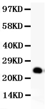
| Description | Rabbit IgG polyclonal antibody for Glial cell line-derived neurotrophic factor(GDNF) detection. Tested with WB in Human;Rat. |
| Reconstitution | Add 0.2ml of distilled water will yield a concentration of 500ug/ml. |
| Gene ID | 2668 |
|---|---|
| Other Names | Glial cell line-derived neurotrophic factor, hGDNF, Astrocyte-derived trophic factor, ATF, GDNF |
| Calculated MW | 23720 MW KDa |
| Application Details | Western blot, 0.1-0.5 µg/ml, Rat, Human |
| Subcellular Localization | Secreted . |
| Tissue Specificity | In the brain, predominantly expressed in the striatum with highest levels in the caudate and lowest in the putamen. Isoform 2 is absent from most tissues except for low levels in intestine and kidney. Highest expression of isoform 3 is found in pancreatic islets. Isoform 5 is expressed at very low levels in putamen, nucleus accumbens, prefrontal cortex, amygdala, hypothalamus and intestine. Isoform 3 is up-regulated in the middle temporal gyrus of Alzheimer disease patients while isoform 2 shows no change. . |
| Protein Name | Glial cell line-derived neurotrophic factor |
| Contents | Each vial contains 5mg BSA, 0.9mg NaCl, 0.2mg Na2HPO4, 0.05mg NaN3. |
| Immunogen | E.coli-derived human GDNF recombinant protein (Position: S78-I211). Human GDNF shares 93% amino acid (aa) sequence identity with both mouse and rat GDNF. |
| Purification | Immunogen affinity purified. |
| Cross Reactivity | No cross reactivity with other proteins |
| Storage | At -20˚C for one year. After r˚Constitution, at 4˚C for one month. It˚Can also be aliquotted and stored frozen at -20˚C for a longer time.Avoid repeated freezing and thawing. |
| Sequence Similarities | Belongs to the TGF-beta family. GDNF subfamily. |
| Name | GDNF |
|---|---|
| Function | Neurotrophic factor that enhances survival and morphological differentiation of dopaminergic neurons and increases their high- affinity dopamine uptake (PubMed:8493557). Acts by binding to its coreceptor, GFRA1, leading to autophosphorylation and activation of the RET receptor (PubMed:10829012, PubMed:25242331, PubMed:31535977). Involved in the development of the neural crest (PubMed:15242795). |
| Cellular Location | Secreted |
| Tissue Location | In the brain, predominantly expressed in the striatum with highest levels in the caudate and lowest in the putamen Isoform 2 is absent from most tissues except for low levels in intestine and kidney. Highest expression of isoform 3 is found in pancreatic islets. Isoform 5 is expressed at very low levels in putamen, nucleus accumbens, prefrontal cortex, amygdala, hypothalamus and intestine. Isoform 3 is up-regulated in the middle temporal gyrus of Alzheimer disease patients while isoform 2 shows no change |

Thousands of laboratories across the world have published research that depended on the performance of antibodies from Abcepta to advance their research. Check out links to articles that cite our products in major peer-reviewed journals, organized by research category.
info@abcepta.com, and receive a free "I Love Antibodies" mug.

Provided below are standard protocols that you may find useful for product applications.
Background
Glial cell line-derived neurotrophic factor(GDNF) is a glycosylated, disulfide-bonded homodimer that is a distantly related member of the transforming growth factor-beta superfamily. GDNF is also a potent neurotrophic factor that promotes the survival of dopaminergic neurones in cultures including embryonic neuronal cultures. In addition to its potential role in the differentiation and survival of central nervous system neurons, it has profound effects on kidney organogenesis and the development of the peripheral nervous system. GDNF may have utility in the treatment of Parkinson's disease, which is marked by progressive degeneration of midbrain dopaminergic neurons.
If you have used an Abcepta product and would like to share how it has performed, please click on the "Submit Review" button and provide the requested information. Our staff will examine and post your review and contact you if needed.
If you have any additional inquiries please email technical services at tech@abcepta.com.
Ordering Information
Other Products
Shipping Information
