


 Foundational characteristics of cancer include proliferation, angiogenesis, migration, evasion of apoptosis, and cellular immortality. Find key markers for these cellular processes and antibodies to detect them.
 The SUMOplot™ Analysis Program predicts and scores sumoylation sites in your protein. SUMOylation is a post-translational modification involved in various cellular processes, such as nuclear-cytosolic transport, transcriptional regulation, apoptosis, protein stability, response to stress, and progression through the cell cycle.
 The Autophagy Receptor Motif Plotter predicts and scores autophagy receptor binding sites in your protein. Identifying proteins connected to this pathway is critical to understanding the role of autophagy in physiological as well as pathological processes such as development, differentiation, neurodegenerative diseases, stress, infection, and cancer.
Anti-CD63 Picoband Antibody
- SPECIFICATION
- CITATIONS
- PROTOCOLS
- BACKGROUND

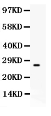
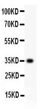
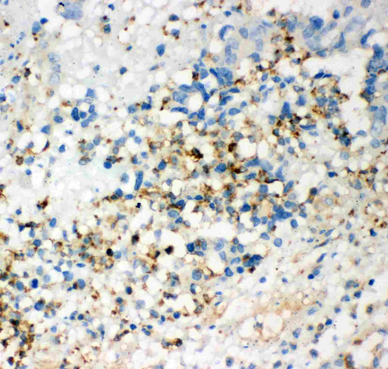
Application
| WB, IHC-P |
|---|---|
| Primary Accession | P08962 |
| Host | Rabbit |
| Reactivity | Human |
| Clonality | Polyclonal |
| Format | Lyophilized |
| Description | Rabbit IgG polyclonal antibody for CD63 antigen(CD63) detection. Tested with WB, IHC-P in Human. |
| Reconstitution | Add 0.2ml of distilled water will yield a concentration of 500ug/ml. |
| Gene ID | 967 |
|---|---|
| Other Names | CD63 antigen, Granulophysin, Lysosomal-associated membrane protein 3, LAMP-3, Melanoma-associated antigen ME491, OMA81H, Ocular melanoma-associated antigen, Tetraspanin-30, Tspan-30, CD63, CD63, MLA1, TSPAN30 |
| Calculated MW | 25637 MW KDa |
| Application Details | Immunohistochemistry(Paraffin-embedded Section), 0.5-1 µg/ml, Human, By Heat Western blot, 0.1-0.5 µg/ml, Human |
| Subcellular Localization | Cell membrane; Multi-pass membrane protein. Lysosome membrane; Multi-pass membrane protein. Late endosome membrane; Multi-pass membrane protein. Endosome, multivesicular body. Melanosome. Also found in Weibel-Palade bodies of endothelial cells. Located in platelet dense granules. Detected in a subset of pre-melanosomes. Detected on intralumenal vesicles (ILVs) within multivesicular bodies. |
| Tissue Specificity | Detected in platelets (at protein level). Dysplastic nevi, radial growth phase primary melanomas, hematopoietic cells, tissue macrophages. . |
| Protein Name | CD63 antigen |
| Contents | Each vial contains 5mg BSA, 0.9mg NaCl, 0.2mg Na2HPO4, 0.05mg NaN3. |
| Immunogen | E.coli-derived human CD63 recombinant protein (Position: E97-M238). Human CD63 shares 74% and 73% amino acid (aa) sequence identity with mouse and rat CD63, respectively. |
| Purification | Immunogen affinity purified. |
| Cross Reactivity | No cross reactivity with other proteins |
| Storage | At -20˚C for one year. After r˚Constitution, at 4˚C for one month. It˚Can also be aliquotted and stored frozen at -20˚C for a longer time.Avoid repeated freezing and thawing. |
| Name | CD63 |
|---|---|
| Synonyms | MLA1, TSPAN30 |
| Function | Functions as a cell surface receptor for TIMP1 and plays a role in the activation of cellular signaling cascades. Plays a role in the activation of ITGB1 and integrin signaling, leading to the activation of AKT, FAK/PTK2 and MAP kinases. Promotes cell survival, reorganization of the actin cytoskeleton, cell adhesion, spreading and migration, via its role in the activation of AKT and FAK/PTK2. Plays a role in VEGFA signaling via its role in regulating the internalization of KDR/VEGFR2. Plays a role in intracellular vesicular transport processes, and is required for normal trafficking of the PMEL luminal domain that is essential for the development and maturation of melanocytes. Plays a role in the adhesion of leukocytes onto endothelial cells via its role in the regulation of SELP trafficking. May play a role in mast cell degranulation in response to Ms4a2/FceRI stimulation, but not in mast cell degranulation in response to other stimuli. |
| Cellular Location | Cell membrane; Multi-pass membrane protein. Lysosome membrane; Multi-pass membrane protein. Late endosome membrane; Multi-pass membrane protein. Endosome, multivesicular body. Melanosome. Secreted, extracellular exosome. Cell surface. Note=Also found in Weibel-Palade bodies of endothelial cells (PubMed:10793155). Located in platelet dense granules (PubMed:7682577). Detected in a subset of pre-melanosomes Detected on intralumenal vesicles (ILVs) within multivesicular bodies (PubMed:21962903). |
| Tissue Location | Detected in platelets (at protein level). Dysplastic nevi, radial growth phase primary melanomas, hematopoietic cells, tissue macrophages. |

Thousands of laboratories across the world have published research that depended on the performance of antibodies from Abcepta to advance their research. Check out links to articles that cite our products in major peer-reviewed journals, organized by research category.
info@abcepta.com, and receive a free "I Love Antibodies" mug.

Provided below are standard protocols that you may find useful for product applications.
Background
CD63 antigen is a protein that in humans is encoded by the CD63 gene. The protein encoded by this gene is a member of the transmembrane 4 superfamily, also known as the tetraspanin family. Most of these members are cell-surface proteins that are characterized by the presence of four hydrophobic domains. It is mapped to 12q13.2. CD63 is mainly associated with membranes of intracellular vesicles, although cell surface expression may be induced. The proteins mediate signal transduction events that play a role in the regulation of cell development, activation, growth and motility. This encoded protein is a cell surface glycoprotein that is known to complex with integrins. It may function as a blood platelet activation marker. Deficiency of this protein is associated with Hermansky-Pudlak syndrome. Also this gene has been associated with tumor progression.
If you have used an Abcepta product and would like to share how it has performed, please click on the "Submit Review" button and provide the requested information. Our staff will examine and post your review and contact you if needed.
If you have any additional inquiries please email technical services at tech@abcepta.com.
Ordering Information
Other Products
Shipping Information
