


 Foundational characteristics of cancer include proliferation, angiogenesis, migration, evasion of apoptosis, and cellular immortality. Find key markers for these cellular processes and antibodies to detect them.
 The SUMOplot™ Analysis Program predicts and scores sumoylation sites in your protein. SUMOylation is a post-translational modification involved in various cellular processes, such as nuclear-cytosolic transport, transcriptional regulation, apoptosis, protein stability, response to stress, and progression through the cell cycle.
 The Autophagy Receptor Motif Plotter predicts and scores autophagy receptor binding sites in your protein. Identifying proteins connected to this pathway is critical to understanding the role of autophagy in physiological as well as pathological processes such as development, differentiation, neurodegenerative diseases, stress, infection, and cancer.
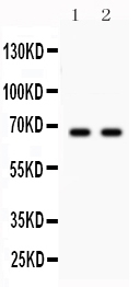
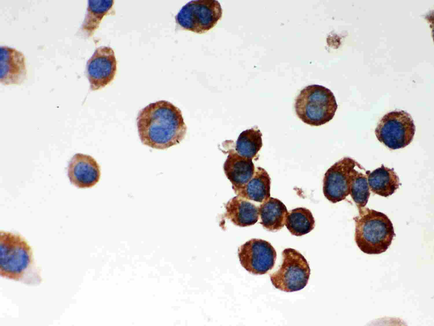
Anti-Parkin Picoband Antibody
- SPECIFICATION
- CITATIONS
- PROTOCOLS
- BACKGROUND

Application
| WB, IHC-P, ICC |
|---|---|
| Primary Accession | O60260 |
| Host | Rabbit |
| Reactivity | Human, Mouse, Rat |
| Clonality | Polyclonal |
| Format | Lyophilized |
| Description | Rabbit IgG polyclonal antibody for E3 ubiquitin-protein ligase parkin(PARK2) detection. Tested with WB, IHC-P, ICC in Human;Mouse;Rat. |
| Reconstitution | Add 0.2ml of distilled water will yield a concentration of 500ug/ml. |
| Gene ID | 5071 |
|---|---|
| Other Names | E3 ubiquitin-protein ligase parkin, Parkin, 2.3.2.-, Parkinson juvenile disease protein 2, Parkinson disease protein 2, PARK2, PRKN |
| Calculated MW | 51641 MW KDa |
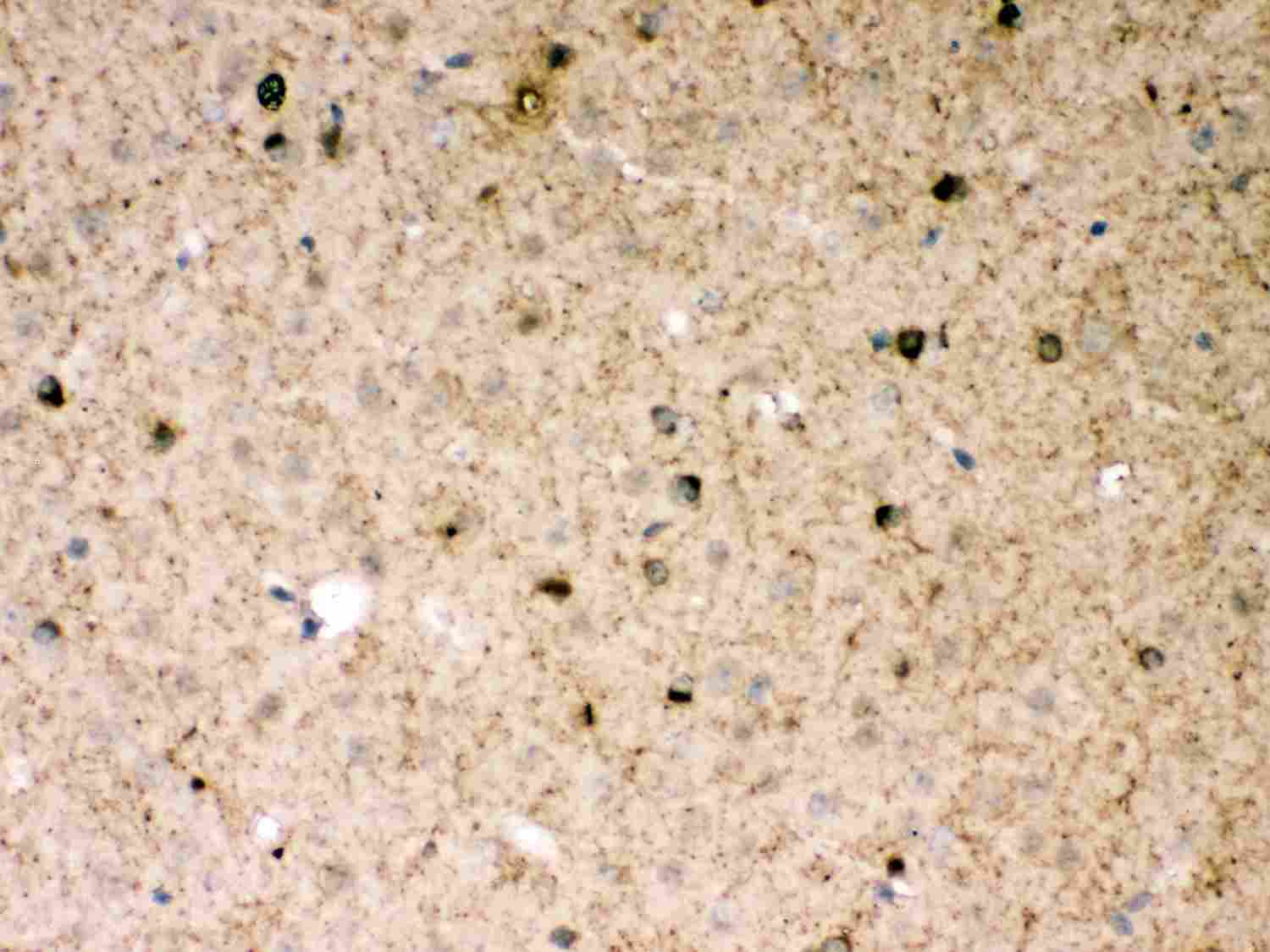
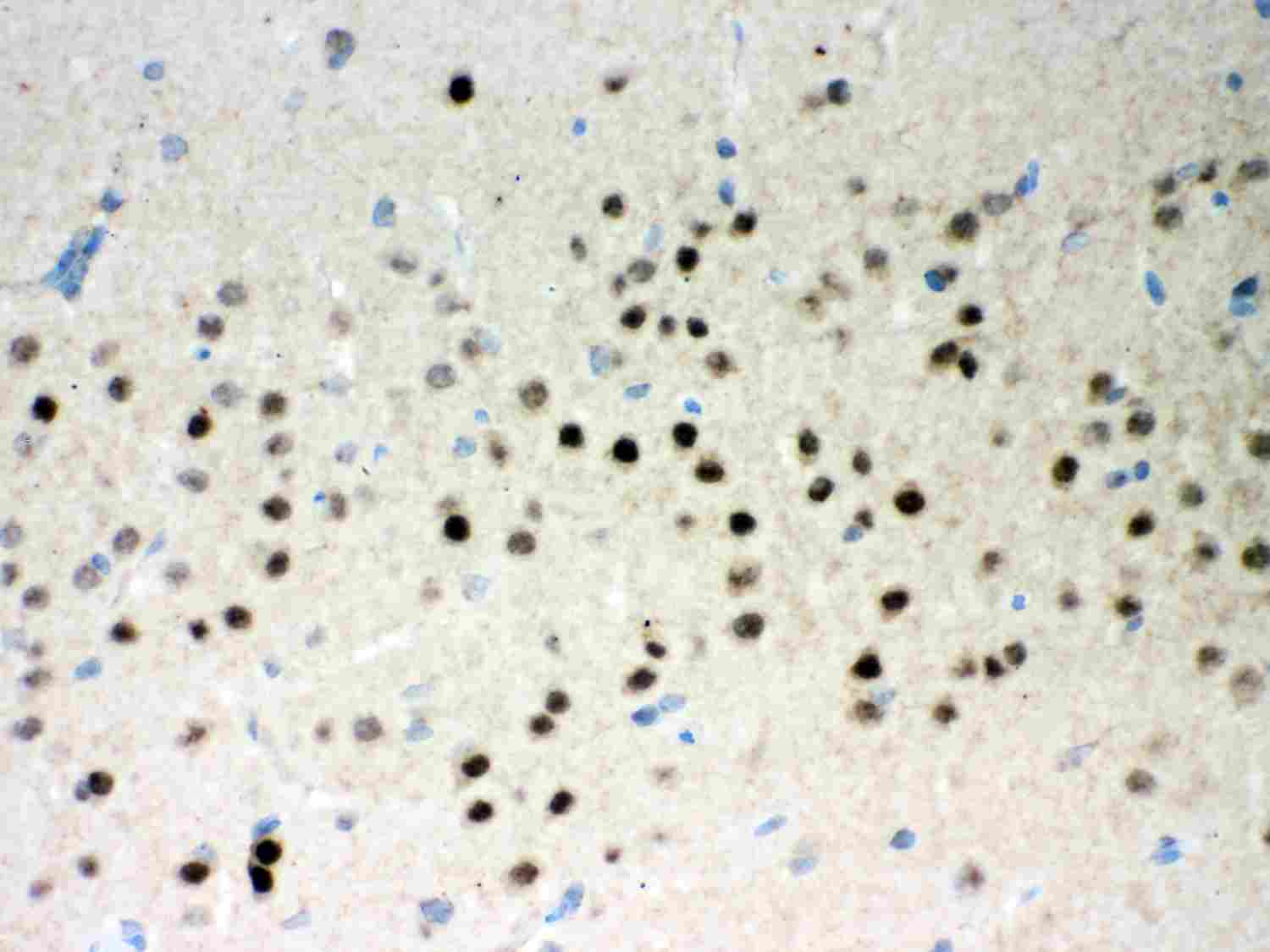
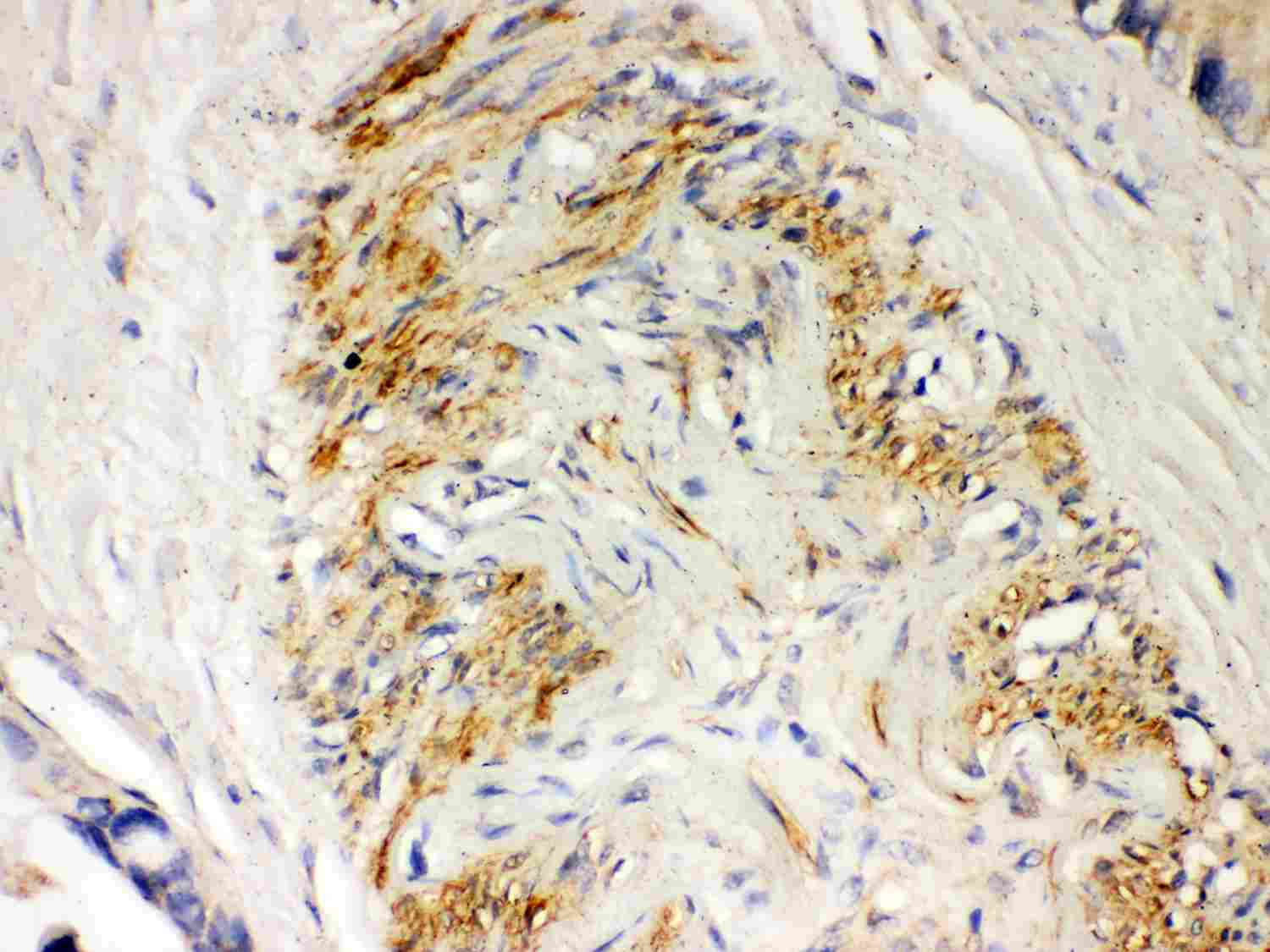
| Application Details | Immunocytochemistry , 0.5-1 µg/ml, Mouse, - Immunohistochemistry(Paraffin-embedded Section), 0.5-1 µg/ml, Human, Mouse, Rat, By Heat Western blot, 0.1-0.5 µg/ml, Human, Mouse, Rat |
| Subcellular Localization | Cytoplasm, cytosol . Nucleus. Endoplasmic reticulum. Mitochondrion . Mainly localizes in the cytosol. Co-localizes with SYT11 in neutrites. Co-localizes with SNCAIP in brainstem Lewy bodies. Mitochondrial localization gradually increases with cellular growth. Also relocates to dysfunctional mitochondria that have lost the mitochondrial membrane potential; recruitment to mitochondria is PINK1- dependent. |
| Tissue Specificity | Highly expressed in the brain including the substantia nigra. Expressed in heart, testis and skeletal muscle. Expression is down-regulated or absent in tumor biopsies, and absent in the brain of PARK2 patients. Overexpression protects dopamine neurons from kainate-mediated apoptosis. Found in serum (at protein level). . |
| Protein Name | E3 ubiquitin-protein ligase parkin |
| Contents | Each vial contains 5mg BSA, 0.9mg NaCl, 0.2mg Na2HPO4, 0.05mg NaN3. |
| Immunogen | E.coli-derived human Parkin recombinant protein (Position: I23-K416). Human Parkin shares 82% and 84% amino acid (aa) sequence identity with mouse and rat Parkin, respectively. |
| Purification | Immunogen affinity purified. |
| Cross Reactivity | No cross reactivity with other proteins |
| Storage | At -20˚C for one year. After r˚Constitution, at 4˚C for one month. It˚Can also be aliquotted and stored frozen at -20˚C for a longer time.Avoid repeated freezing and thawing. |
| Sequence Similarities | Belongs to the RBR family. Parkin subfamily. |
| Name | PRKN (HGNC:8607) |
|---|---|
| Synonyms | PARK2 |
| Function | Functions within a multiprotein E3 ubiquitin ligase complex, catalyzing the covalent attachment of ubiquitin moieties onto substrate proteins (PubMed:10888878, PubMed:10973942, PubMed:11431533, PubMed:12150907, PubMed:12628165, PubMed:15105460, PubMed:16135753, PubMed:21376232, PubMed:21532592, PubMed:22396657, PubMed:23620051, PubMed:23754282, PubMed:24660806, PubMed:24751536, PubMed:29311685, PubMed:32047033). Substrates include SYT11 and VDAC1 (PubMed:29311685, PubMed:32047033). Other substrates are BCL2, CCNE1, GPR37, RHOT1/MIRO1, MFN1, MFN2, STUB1, SNCAIP, SEPTIN5, TOMM20, USP30, ZNF746, MIRO1 and AIMP2 (PubMed:10888878, PubMed:10973942, PubMed:11431533, PubMed:12150907, PubMed:12628165, PubMed:15105460, PubMed:16135753, PubMed:21376232, PubMed:21532592, PubMed:22396657, PubMed:23620051, PubMed:23754282, PubMed:24660806, PubMed:24751536). Mediates monoubiquitination as well as 'Lys-6', 'Lys-11', 'Lys-48'-linked and 'Lys-63'-linked polyubiquitination of substrates depending on the context (PubMed:19229105, PubMed:20889974, PubMed:25474007, PubMed:25621951, PubMed:32047033). Participates in the removal and/or detoxification of abnormally folded or damaged protein by mediating 'Lys-63'-linked polyubiquitination of misfolded proteins such as PARK7: 'Lys-63'-linked polyubiquitinated misfolded proteins are then recognized by HDAC6, leading to their recruitment to aggresomes, followed by degradation (PubMed:17846173, PubMed:19229105). Mediates 'Lys-63'-linked polyubiquitination of a 22 kDa O-linked glycosylated isoform of SNCAIP, possibly playing a role in Lewy-body formation (PubMed:11431533, PubMed:11590439, PubMed:15105460, PubMed:15728840, PubMed:19229105). Mediates monoubiquitination of BCL2, thereby acting as a positive regulator of autophagy (PubMed:20889974). Protects against mitochondrial dysfunction during cellular stress, by acting downstream of PINK1 to coordinate mitochondrial quality control mechanisms that remove and replace dysfunctional mitochondrial components (PubMed:11439185, PubMed:18957282, PubMed:19029340, PubMed:19966284, PubMed:21376232, PubMed:22082830, PubMed:22396657, PubMed:23620051, PubMed:23933751, PubMed:24660806, PubMed:24784582, PubMed:24896179, PubMed:25474007, PubMed:25527291, PubMed:32047033). Depending on the severity of mitochondrial damage and/or dysfunction, activity ranges from preventing apoptosis and stimulating mitochondrial biogenesis to regulating mitochondrial dynamics and eliminating severely damaged mitochondria via mitophagy (PubMed:11439185, PubMed:19029340, PubMed:19801972, PubMed:19966284, PubMed:21376232, PubMed:22082830, PubMed:22396657, PubMed:23620051, PubMed:23685073, PubMed:23933751, PubMed:24896179, PubMed:25527291, PubMed:32047033, PubMed:33499712). Activation and recruitment onto the outer membrane of damaged/dysfunctional mitochondria (OMM) requires PINK1-mediated phosphorylation of both PRKN and ubiquitin (PubMed:24660806, PubMed:24784582, PubMed:25474007, PubMed:25527291). After mitochondrial damage, functions with PINK1 to mediate the decision between mitophagy or preventing apoptosis by inducing either the poly- or monoubiquitination of VDAC1, respectively; polyubiquitination of VDAC1 promotes mitophagy, while monoubiquitination of VDAC1 decreases mitochondrial calcium influx which ultimately inhibits apoptosis (PubMed:27534820, PubMed:32047033). When cellular stress results in irreversible mitochondrial damage, promotes the autophagic degradation of dysfunctional depolarized mitochondria (mitophagy) by promoting the ubiquitination of mitochondrial proteins such as TOMM20, RHOT1/MIRO1, MFN1 and USP30 (PubMed:19029340, PubMed:19966284, PubMed:21753002, PubMed:22396657, PubMed:23620051, PubMed:23685073, PubMed:23933751, PubMed:24896179, PubMed:25527291). Preferentially assembles 'Lys-6'-, 'Lys-11'- and 'Lys-63'-linked polyubiquitin chains, leading to mitophagy (PubMed:25621951, PubMed:32047033). The PINK1-PRKN pathway also promotes fission of damaged mitochondria by PINK1-mediated phosphorylation which promotes the PRKN-dependent degradation of mitochondrial proteins involved in fission such as MFN2 (PubMed:23620051). This prevents the refusion of unhealthy mitochondria with the mitochondrial network or initiates mitochondrial fragmentation facilitating their later engulfment by autophagosomes (PubMed:23620051). Regulates motility of damaged mitochondria via the ubiquitination and subsequent degradation of MIRO1 and MIRO2; in motor neurons, this likely inhibits mitochondrial intracellular anterograde transport along the axons which probably increases the chance of the mitochondria undergoing mitophagy in the soma (PubMed:22396657). Involved in mitochondrial biogenesis via the 'Lys-48'-linked polyubiquitination of transcriptional repressor ZNF746/PARIS which leads to its subsequent proteasomal degradation and allows activation of the transcription factor PPARGC1A (PubMed:21376232). Limits the production of reactive oxygen species (ROS) (PubMed:18541373). Regulates cyclin-E during neuronal apoptosis (PubMed:12628165). In collaboration with CHPF isoform 2, may enhance cell viability and protect cells from oxidative stress (PubMed:22082830). Independently of its ubiquitin ligase activity, protects from apoptosis by the transcriptional repression of p53/TP53 (PubMed:19801972). May protect neurons against alpha synuclein toxicity, proteasomal dysfunction, GPR37 accumulation, and kainate-induced excitotoxicity (PubMed:11439185). May play a role in controlling neurotransmitter trafficking at the presynaptic terminal and in calcium-dependent exocytosis. May represent a tumor suppressor gene (PubMed:12719539). |
| Cellular Location | Cytoplasm, cytosol. Nucleus. Endoplasmic reticulum. Mitochondrion. Mitochondrion outer membrane {ECO:0000250|UniProtKB:Q9WVS6}. Cell projection, neuron projection. Postsynaptic density {ECO:0000250|UniProtKB:Q9WVS6}. Presynapse {ECO:0000250|UniProtKB:Q9WVS6}. Note=Mainly localizes in the cytosol (PubMed:19029340, PubMed:19229105). Co-localizes with SYT11 in neutrites (PubMed:12925569). Co-localizes with SNCAIP in brainstem Lewy bodies (PubMed:10319893, PubMed:11431533). Translocates to dysfunctional mitochondria that have lost the mitochondrial membrane potential; recruitment to mitochondria is PINK1-dependent (PubMed:18957282, PubMed:19966284, PubMed:23620051, PubMed:24898855) Mitochondrial localization also gradually increases with cellular growth (PubMed:22082830). |
| Tissue Location | Highly expressed in the brain including the substantia nigra (PubMed:19501131, PubMed:9560156). Expressed in heart, testis and skeletal muscle (PubMed:9560156). Expression is down- regulated or absent in tumor biopsies, and absent in the brain of PARK2 patients (PubMed:12719539, PubMed:14614460). Overexpression protects dopamine neurons from kainate-mediated apoptosis (PubMed:12628165) Found in serum (at protein level) (PubMed:19501131) |

Thousands of laboratories across the world have published research that depended on the performance of antibodies from Abcepta to advance their research. Check out links to articles that cite our products in major peer-reviewed journals, organized by research category.
info@abcepta.com, and receive a free "I Love Antibodies" mug.

Provided below are standard protocols that you may find useful for product applications.
Background
Parkin is a RING domain-containing E3 ubiquitin ligase involved in proteasome-dependent degradation of proteins. It is mapped to 6q26. This gene is important for mitochondrial quality control by lysosome-dependent degradation of damaged mitochondria through autophagy, or mitophagy. Parkin is expressed in neuronal processes and cell bodies of neurons, but not glial cells, in the midbrain, basal ganglia, cerebral cortex, and cerebellum. Parkin assimilated with actin filaments, suggesting that it is a cytoskeletal-associated protein. Parkin is identified as a transcriptional repressor of p53 independent of its ubiquitin ligase function. It also has been found that parkin was associated physically with mitochondrial DNA (mtDNA) in proliferating as well as in differentiated SH-SY5Y neuroblastoma cells.
If you have used an Abcepta product and would like to share how it has performed, please click on the "Submit Review" button and provide the requested information. Our staff will examine and post your review and contact you if needed.
If you have any additional inquiries please email technical services at tech@abcepta.com.
Ordering Information
Other Products
Shipping Information
