


 Foundational characteristics of cancer include proliferation, angiogenesis, migration, evasion of apoptosis, and cellular immortality. Find key markers for these cellular processes and antibodies to detect them.
 The SUMOplot™ Analysis Program predicts and scores sumoylation sites in your protein. SUMOylation is a post-translational modification involved in various cellular processes, such as nuclear-cytosolic transport, transcriptional regulation, apoptosis, protein stability, response to stress, and progression through the cell cycle.
 The Autophagy Receptor Motif Plotter predicts and scores autophagy receptor binding sites in your protein. Identifying proteins connected to this pathway is critical to understanding the role of autophagy in physiological as well as pathological processes such as development, differentiation, neurodegenerative diseases, stress, infection, and cancer.
Anti-Peroxiredoxin 3 Picoband Antibody
- SPECIFICATION
- CITATIONS
- PROTOCOLS
- BACKGROUND

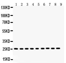
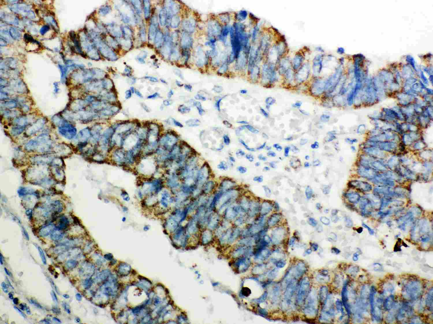
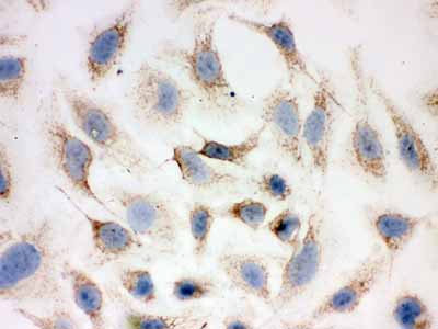
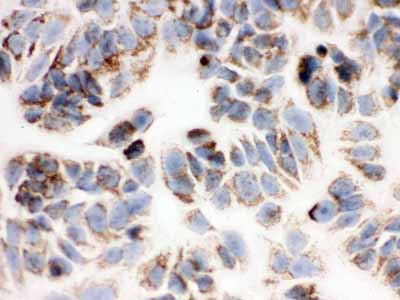
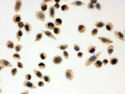
Application
| WB, IHC-P, ICC |
|---|---|
| Primary Accession | P30048 |
| Host | Rabbit |
| Reactivity | Human, Mouse, Rat |
| Clonality | Polyclonal |
| Format | Lyophilized |
| Description | Rabbit IgG polyclonal antibody for Thioredoxin-dependent peroxide reductase, mitochondrial(PRDX3) detection. Tested with WB, IHC-P, ICC in Human;Mouse;Rat. |
| Reconstitution | Add 0.2ml of distilled water will yield a concentration of 500ug/ml. |
| Gene ID | 10935 |
|---|---|
| Other Names | Thioredoxin-dependent peroxide reductase, mitochondrial, 1.11.1.15, Antioxidant protein 1, AOP-1, HBC189, Peroxiredoxin III, Prx-III, Peroxiredoxin-3, Protein MER5 homolog, PRDX3, AOP1 |
| Calculated MW | 27693 MW KDa |
| Application Details | Immunohistochemistry(Paraffin-embedded Section), 0.5-1 µg/ml, By Heat Immunocytochemistry, 0.5-1 µg/ml Western blot, 0.1-0.5 µg/ml |
| Subcellular Localization | Mitochondrion. |
| Protein Name | Thioredoxin-dependent peroxide reductase, mitochondrial |
| Contents | Each vial contains 5mg BSA, 0.9mg NaCl, 0.2mg Na2HPO4, 0.05mg NaN3. |
| Immunogen | E.coli-derived human Peroxiredoxin 3 recombinant protein (Position: T110-Q256). Human Peroxiredoxin 3 shares 93% amino acid (aa) sequence identity with both mouse and rat Peroxiredoxin 3. |
| Purification | Immunogen affinity purified. |
| Cross Reactivity | No cross reactivity with other proteins. |
| Storage | At -20˚C for one year. After r˚Constitution, at 4˚C for one month. It˚Can also be aliquotted and stored frozen at -20˚C for a longer time.Avoid repeated freezing and thawing. |
| Sequence Similarities | Belongs to the AhpC/TSA family. |
| Name | PRDX3 |
|---|---|
| Synonyms | AOP1 |
| Function | Thiol-specific peroxidase that catalyzes the reduction of hydrogen peroxide and organic hydroperoxides to water and alcohols, respectively. Plays a role in cell protection against oxidative stress by detoxifying peroxides (PubMed:17707404, PubMed:29438714, PubMed:33889951, PubMed:7733872). Acts synergistically with MAP3K13 to regulate the activation of NF-kappa-B in the cytosol (PubMed:12492477). Required for the maintenance of physical strength (By similarity). |
| Cellular Location | Mitochondrion. Cytoplasm. Early endosome. Note=Localizes to early endosomes in a RPS6KC1-dependent manner. |

Thousands of laboratories across the world have published research that depended on the performance of antibodies from Abcepta to advance their research. Check out links to articles that cite our products in major peer-reviewed journals, organized by research category.
info@abcepta.com, and receive a free "I Love Antibodies" mug.

Provided below are standard protocols that you may find useful for product applications.
Background
PRDX3(peroxiredoxin 3) also known as AOP-1, MER5, SP-22 or PRX3, is localized exclusively in mitochondria. The deduced 256-amino acid human AOP1 protein shares 86% amino acid sequence similarity with mouse Aop1, and significant similarity with both the human proliferation-associated gene A product and the mouse stress-induced peritoneal macrophage protein Msp23. The PRDX3 gene is mapped on 10q26.11. Expression of PRDX3 is induced by MYC and is reduced in c-myc -/- cells. Chromatin immunoprecipitation analysis spanning the entire PRDX3 genomic sequence revealed that MYC binds preferentially to a 930-bp region surrounding exon 1. Results using mitochondria-specific fluorescent probes demonstrated that PRDX3 is essential for maintaining mitochondrial mass and membrane potential in transformed rat and human cells. These data provided evidence that PRDX3 is a MYC target gene that is required to maintain normal mitochondrial function.
If you have used an Abcepta product and would like to share how it has performed, please click on the "Submit Review" button and provide the requested information. Our staff will examine and post your review and contact you if needed.
If you have any additional inquiries please email technical services at tech@abcepta.com.
Ordering Information
Other Products
Shipping Information
