


 Foundational characteristics of cancer include proliferation, angiogenesis, migration, evasion of apoptosis, and cellular immortality. Find key markers for these cellular processes and antibodies to detect them.
 The SUMOplot™ Analysis Program predicts and scores sumoylation sites in your protein. SUMOylation is a post-translational modification involved in various cellular processes, such as nuclear-cytosolic transport, transcriptional regulation, apoptosis, protein stability, response to stress, and progression through the cell cycle.
 The Autophagy Receptor Motif Plotter predicts and scores autophagy receptor binding sites in your protein. Identifying proteins connected to this pathway is critical to understanding the role of autophagy in physiological as well as pathological processes such as development, differentiation, neurodegenerative diseases, stress, infection, and cancer.
Anti-CD55 Picoband Antibody
- SPECIFICATION
- CITATIONS
- PROTOCOLS
- BACKGROUND

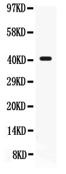
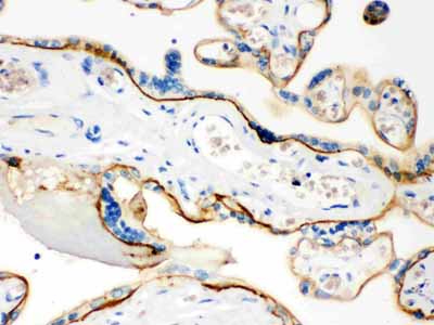
Application
| WB, IHC-P |
|---|---|
| Primary Accession | P08174 |
| Host | Rabbit |
| Reactivity | Human, Mouse |
| Clonality | Polyclonal |
| Format | Lyophilized |
| Description | Rabbit IgG polyclonal antibody for Complement decay-accelerating factor(CD55) detection. Tested with WB, IHC-P in Human;Mouse. |
| Reconstitution | Add 0.2ml of distilled water will yield a concentration of 500ug/ml. |
| Gene ID | 1604 |
|---|---|
| Other Names | Complement decay-accelerating factor, CD55, CD55, CR, DAF |
| Calculated MW | 41400 MW KDa |
| Application Details | Immunohistochemistry(Paraffin-embedded Section), 0.5-1 µg/ml, Human, By Heat Western blot, 0.1-0.5 µg/ml, Human, Mouse |
| Subcellular Localization | Isoform 1: Cell membrane; Single-pass type I membrane protein. |
| Tissue Specificity | Expressed on the plasma membranes of all cell types that are in intimate contact with plasma complement proteins. It is also found on the surfaces of epithelial cells lining extracellular compartments, and variants of the molecule are present in body fluids and in extracellular matrix. |
| Protein Name | Complement decay-accelerating factor |
| Contents | Each vial contains 5mg BSA, 0.9mg NaCl, 0.2mg Na2HPO4, 0.05mg NaN3. |
| Immunogen | E.coli-derived human CD55 recombinant protein (Position: D35-K347). Human CD55 shares 49.1% amino acid (aa) sequence identity with mouse CD55. |
| Purification | Immunogen affinity purified. |
| Cross Reactivity | No cross reactivity with other proteins |
| Storage | At -20˚C for one year. After r˚Constitution, at 4˚C for one month. It˚Can also be aliquotted and stored frozen at -20˚C for a longer time.Avoid repeated freezing and thawing. |
| Sequence Similarities | Belongs to the receptors of complement activation (RCA) family. |
| Name | CD55 |
|---|---|
| Synonyms | CR, DAF |
| Function | This protein recognizes C4b and C3b fragments that condense with cell-surface hydroxyl or amino groups when nascent C4b and C3b are locally generated during C4 and c3 activation. Interaction of daf with cell-associated C4b and C3b polypeptides interferes with their ability to catalyze the conversion of C2 and factor B to enzymatically active C2a and Bb and thereby prevents the formation of C4b2a and C3bBb, the amplification convertases of the complement cascade (PubMed:7525274). Inhibits complement activation by destabilizing and preventing the formation of C3 and C5 convertases, which prevents complement damage (PubMed:28657829). |
| Cellular Location | [Isoform 1]: Cell membrane; Single-pass type I membrane protein [Isoform 3]: Secreted [Isoform 5]: Secreted [Isoform 7]: Cell membrane; Lipid-anchor, GPI-anchor |
| Tissue Location | Expressed on the plasma membranes of all cell types that are in intimate contact with plasma complement proteins. It is also found on the surfaces of epithelial cells lining extracellular compartments, and variants of the molecule are present in body fluids and in extracellular matrix |

Thousands of laboratories across the world have published research that depended on the performance of antibodies from Abcepta to advance their research. Check out links to articles that cite our products in major peer-reviewed journals, organized by research category.
info@abcepta.com, and receive a free "I Love Antibodies" mug.

Provided below are standard protocols that you may find useful for product applications.
Background
Complement decay-accelerating factor, also known as CD55 or DAF, is a protein that, in humans, is encoded by the CD55 gene. This gene encodes a glycoprotein involved in the regulation of the complement cascade. Binding of the encoded protein to complement proteins accelerates their decay, thereby disrupting the cascade and preventing damage to host cells. Antigens present on this protein constitute the Cromer blood group system (CROM). Alternative splicing results in multiple transcript variants. The predominant transcript variant encodes a membrane-bound protein, but alternatively spliced transcripts may produce soluble proteins.
If you have used an Abcepta product and would like to share how it has performed, please click on the "Submit Review" button and provide the requested information. Our staff will examine and post your review and contact you if needed.
If you have any additional inquiries please email technical services at tech@abcepta.com.
Ordering Information
Other Products
Shipping Information
