


 Foundational characteristics of cancer include proliferation, angiogenesis, migration, evasion of apoptosis, and cellular immortality. Find key markers for these cellular processes and antibodies to detect them.
 The SUMOplot™ Analysis Program predicts and scores sumoylation sites in your protein. SUMOylation is a post-translational modification involved in various cellular processes, such as nuclear-cytosolic transport, transcriptional regulation, apoptosis, protein stability, response to stress, and progression through the cell cycle.
 The Autophagy Receptor Motif Plotter predicts and scores autophagy receptor binding sites in your protein. Identifying proteins connected to this pathway is critical to understanding the role of autophagy in physiological as well as pathological processes such as development, differentiation, neurodegenerative diseases, stress, infection, and cancer.
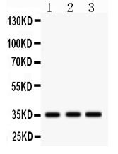
Anti-IGFBP2 Picoband Antibody
- SPECIFICATION
- CITATIONS
- PROTOCOLS
- BACKGROUND

Application
| WB |
|---|---|
| Primary Accession | P18065 |
| Host | Rabbit |
| Reactivity | Human, Rat |
| Clonality | Polyclonal |
| Format | Lyophilized |
| Description | Rabbit IgG polyclonal antibody for Insulin-like growth factor-binding protein 2(IGFBP2) detection. Tested with WB in Human;Rat. |
| Reconstitution | Add 0.2ml of distilled water will yield a concentration of 500ug/ml. |
| Gene ID | 3485 |
|---|---|
| Other Names | Insulin-like growth factor-binding protein 2, IBP-2, IGF-binding protein 2, IGFBP-2, IGFBP2, BP2, IBP2 |
| Calculated MW | 34814 MW KDa |
| Application Details | Western blot, 0.1-0.5 µg/ml, Human, Rat |
| Subcellular Localization | Secreted. |
| Protein Name | Insulin-like growth factor-binding protein 2 |
| Contents | Each vial contains 5mg BSA, 0.9mg NaCl, 0.2mg Na2HPO4, 0.05mg NaN3. |
| Immunogen | A synthetic peptide corresponding to a sequence at the C-terminus of human IGFBP2 (228-257aa QQELDQVLERISTMRLPDERGPLEHLYSLH), different from the related mouse and rat sequences by one amino acid. |
| Purification | Immunogen affinity purified. |
| Cross Reactivity | No cross reactivity with other proteins |
| Storage | At -20˚C for one year. After r˚Constitution, at 4˚C for one month. It˚Can also be aliquotted and stored frozen at -20˚C for a longer time.Avoid repeated freezing and thawing. |
| Sequence Similarities | Contains 1 IGFBP N-terminal domain. |
| Name | IGFBP2 |
|---|---|
| Synonyms | BP2, IBP2 |
| Function | Multifunctional protein that plays a critical role in regulating the availability of IGFs such as IGF1 and IGF2 to their receptors and thereby regulates IGF-mediated cellular processes including proliferation, differentiation, and apoptosis in a cell-type specific manner (PubMed:18563800, PubMed:38796567). Functions coordinately with receptor protein tyrosine phosphatase beta/PTPRB and the IGF1 receptor to regulate IGF1-mediated signaling by stimulating the phosphorylation of PTEN leading to its inactivation and AKT1 activation (PubMed:22869525). Plays a positive role in cell migration via interaction with integrin alpha5/ITGA5 through an RGD motif (PubMed:16569642). Additionally, interaction with ITGA5/ITGB1 enhances the adhesion of endothelial progenitor cells to endothelial cells (PubMed:26076738). Upon mitochondrial damage, facilitates apoptosis with ITGA5 of podocytes, and then activates the phosphorylation of focal adhesion kinase (FAK)-mediated mitochondrial injury (PubMed:38796567). |
| Cellular Location | Secreted |

Thousands of laboratories across the world have published research that depended on the performance of antibodies from Abcepta to advance their research. Check out links to articles that cite our products in major peer-reviewed journals, organized by research category.
info@abcepta.com, and receive a free "I Love Antibodies" mug.

Provided below are standard protocols that you may find useful for product applications.
Background
The superfamily of insulin-like growth factor (IGF) binding proteins include the six high-affinity IGF binding proteins (IGFBP) and at least four additional low-affinity binding proteins referred to as IGFBP related proteins (IGFBP-rP). All IGFBP superfamily members are cysteine-rich proteins with conserved cysteine residues, which are clustered in the amino- and carboxy-terminal thirds of the molecule. IGFBPs modulate the biological activities of IGF proteins. Some IGFBPs may also have intrinsic bioactivity that is independent of their ability to bind IGF proteins. Post-translational modifications of IGFBPs, including glycosylation, phosphorylation and proteolysis, have been shown to modify the affinities of the binding proteins to IGF. Human IGFBP-2 cDNA encodes a 328 amino acid (aa) residue precursor protein with a putative 39 aa residue signal peptide that is processed to generate the 289 aa residue mature protein. IGFBP-2 contains an integrin receptor recognition sequence (RGD sequence) but lacks potential N-linked glycosylation sites. During development, IGFBP-2 is expressed in a number of tissues. The highest expression level is found in the central nervous system. In adults, high expression levels are also detected in the central nervous system and in a number of reproductive tissues. IGFBP-2 binds preferentially to IGF II, exhibiting a 2-10 fold higher affinity for IGF II than for IGF I.
If you have used an Abcepta product and would like to share how it has performed, please click on the "Submit Review" button and provide the requested information. Our staff will examine and post your review and contact you if needed.
If you have any additional inquiries please email technical services at tech@abcepta.com.
Ordering Information
Other Products
Shipping Information
