


 Foundational characteristics of cancer include proliferation, angiogenesis, migration, evasion of apoptosis, and cellular immortality. Find key markers for these cellular processes and antibodies to detect them.
 The SUMOplot™ Analysis Program predicts and scores sumoylation sites in your protein. SUMOylation is a post-translational modification involved in various cellular processes, such as nuclear-cytosolic transport, transcriptional regulation, apoptosis, protein stability, response to stress, and progression through the cell cycle.
 The Autophagy Receptor Motif Plotter predicts and scores autophagy receptor binding sites in your protein. Identifying proteins connected to this pathway is critical to understanding the role of autophagy in physiological as well as pathological processes such as development, differentiation, neurodegenerative diseases, stress, infection, and cancer.
Anti-PDPK1 Picoband Antibody
- SPECIFICATION
- CITATIONS
- PROTOCOLS
- BACKGROUND

Application
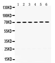
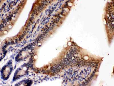
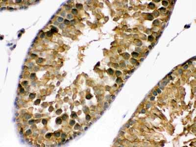
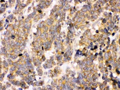
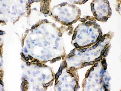
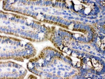
| WB, IHC-P, IHC-F |
|---|---|
| Primary Accession | O15530 |
| Host | Rabbit |
| Reactivity | Human, Mouse, Rat |
| Clonality | Polyclonal |
| Format | Lyophilized |
| Description | Rabbit IgG polyclonal antibody for 3-phosphoinositide-dependent protein kinase 1(PDPK1) detection. Tested with WB, IHC-P, IHC-F in Human;Mouse;Rat. |
| Reconstitution | Add 0.2ml of distilled water will yield a concentration of 500ug/ml. |
| Gene ID | 5170 |
|---|---|
| Other Names | 3-phosphoinositide-dependent protein kinase 1, hPDK1, 2.7.11.1, PDPK1, PDK1 |
| Calculated MW | 63152 MW KDa |
| Application Details | Immunohistochemistry(Paraffin-embedded Section), 0.5-1 µg/ml, By Heat Immunohistochemistry(Frozen Section), 0.5-1 µg/ml Western blot, 0.1-0.5 µg/ml |
| Subcellular Localization | Cytoplasm. Nucleus. Cell membrane; Peripheral membrane protein. Cell junction, focal adhesion. Tyrosine phosphorylation seems to occur only at the cell membrane. Translocates to the cell membrane following insulin stimulation by a mechanism that involves binding to GRB14 and INSR. SRC and HSP90 promote its localization to the cell membrane. Its nuclear localization is dependent on its association with PTPN6 and its phosphorylation at Ser-396. Restricted to the nucleus in neuronal cells while in non-neuronal cells it is found in the cytoplasm. The Ser-241 phosphorylated form is distributed along the perinuclear region in neuronal cells while in non- neuronal cells it is found in both the nucleus and the cytoplasm. IGF1 transiently increases phosphorylation at Ser-241 of neuronal PDPK1, resulting in its translocation to other cellular compartments. The tyrosine-phosphorylated form colocalizes with PTK2B in focal adhesions after angiotensin II stimulation. |
| Tissue Specificity | Appears to be expressed ubiquitously. The Tyr- 9 phosphorylated form is markedly increased in diseased tissue compared with normal tissue from lung, liver, colon and breast. . |
| Protein Name | 3-phosphoinositide-dependent protein kinase 1 |
| Contents | Each vial contains 5mg BSA, 0.9mg NaCl, 0.2mg Na2HPO4, 0.05mg NaN3. |
| Immunogen | A synthetic peptide corresponding to a sequence at the C-terminus of human PDPK1 (524-556aa YLMDPSGNAHKWCRKIQEVWRQRYQSHPDAAVQ), different from the related mouse and rat sequences by two amino acids. |
| Purification | Immunogen affinity purified. |
| Cross Reactivity | No cross reactivity with other proteins. |
| Storage | At -20˚C for one year. After r˚Constitution, at 4˚C for one month. It˚Can also be aliquotted and stored frozen at -20˚C for a longer time.Avoid repeated freezing and thawing. |
| Name | PDPK1 |
|---|---|
| Synonyms | PDK1 |
| Function | Serine/threonine kinase which acts as a master kinase, phosphorylating and activating a subgroup of the AGC family of protein kinases (PubMed:10226025, PubMed:10480933, PubMed:10995762, PubMed:12167717, PubMed:14585963, PubMed:14604990, PubMed:16207722, PubMed:16251192, PubMed:17327236, PubMed:17371830, PubMed:18835241, PubMed:9094314, PubMed:9368760, PubMed:9445476, PubMed:9445477, PubMed:9707564, PubMed:9768361). Its targets include: protein kinase B (PKB/AKT1, PKB/AKT2, PKB/AKT3), p70 ribosomal protein S6 kinase (RPS6KB1), p90 ribosomal protein S6 kinase (RPS6KA1, RPS6KA2 and RPS6KA3), cyclic AMP-dependent protein kinase (PRKACA), protein kinase C (PRKCD and PRKCZ), serum and glucocorticoid-inducible kinase (SGK1, SGK2 and SGK3), p21-activated kinase-1 (PAK1), TSSK3, protein kinase PKN (PKN1 and PKN2) (PubMed:10226025, PubMed:10480933, PubMed:10995762, PubMed:12167717, PubMed:14585963, PubMed:14604990, PubMed:16207722, PubMed:16251192, PubMed:17327236, PubMed:17371830, PubMed:18835241, PubMed:9094314, PubMed:9368760, PubMed:9445476, PubMed:9707564, PubMed:9768361). Plays a central role in the transduction of signals from insulin by providing the activating phosphorylation to PKB/AKT1, thus propagating the signal to downstream targets controlling cell proliferation and survival, as well as glucose and amino acid uptake and storage (PubMed:10226025, PubMed:12167717, PubMed:9094314). Negatively regulates the TGF-beta-induced signaling by: modulating the association of SMAD3 and SMAD7 with TGF-beta receptor, phosphorylating SMAD2, SMAD3, SMAD4 and SMAD7, preventing the nuclear translocation of SMAD3 and SMAD4 and the translocation of SMAD7 from the nucleus to the cytoplasm in response to TGF-beta (PubMed:17327236). Activates PPARG transcriptional activity and promotes adipocyte differentiation (By similarity). Activates the NF-kappa-B pathway via phosphorylation of IKKB (PubMed:16207722). The tyrosine phosphorylated form is crucial for the regulation of focal adhesions by angiotensin II (PubMed:14585963). Controls proliferation, survival, and growth of developing pancreatic cells (By similarity). Participates in the regulation of Ca(2+) entry and Ca(2+)-activated K(+) channels of mast cells (By similarity). Essential for the motility of vascular endothelial cells (ECs) and is involved in the regulation of their chemotaxis (PubMed:17371830). Plays a critical role in cardiac homeostasis by serving as a dual effector for cell survival and beta-adrenergic response (By similarity). Plays an important role during thymocyte development by regulating the expression of key nutrient receptors on the surface of pre-T cells and mediating Notch-induced cell growth and proliferative responses (By similarity). Provides negative feedback inhibition to toll-like receptor-mediated NF-kappa-B activation in macrophages (By similarity). |
| Cellular Location | Cytoplasm. Nucleus. Cell membrane; Peripheral membrane protein. Cell junction, focal adhesion. Note=Tyrosine phosphorylation seems to occur only at the cell membrane. Translocates to the cell membrane following insulin stimulation by a mechanism that involves binding to GRB14 and INSR. SRC and HSP90 promote its localization to the cell membrane. Its nuclear localization is dependent on its association with PTPN6 and its phosphorylation at Ser- 396. Restricted to the nucleus in neuronal cells while in non-neuronal cells it is found in the cytoplasm. The Ser-241 phosphorylated form is distributed along the perinuclear region in neuronal cells while in non-neuronal cells it is found in both the nucleus and the cytoplasm IGF1 transiently increases phosphorylation at Ser-241 of neuronal PDPK1, resulting in its translocation to other cellular compartments The tyrosine-phosphorylated form colocalizes with PTK2B in focal adhesions after angiotensin II stimulation |
| Tissue Location | Appears to be expressed ubiquitously. The Tyr-9 phosphorylated form is markedly increased in diseased tissue compared with normal tissue from lung, liver, colon and breast |

Thousands of laboratories across the world have published research that depended on the performance of antibodies from Abcepta to advance their research. Check out links to articles that cite our products in major peer-reviewed journals, organized by research category.
info@abcepta.com, and receive a free "I Love Antibodies" mug.

Provided below are standard protocols that you may find useful for product applications.
Background
3-phosphoinositide dependent protein kinase-1, also known as PDPK1, is a protein which in humans is encoded by the PDPK1 gene. It is mapped to 16p13.3. PDPK1 is a master kinase, which is crucial for the activation of AKT/PKB and many other AGC kinases including PKC, S6K, SGK. An important role for PDPK1 is in the signalling pathways activated by several growth factors and hormones including insulin signaling. Mice lacking PDPK1 die during early embryonic development, indicating that this enzyme is critical for transmitting the growth-promoting signals necessary for normal mammalian development.
If you have used an Abcepta product and would like to share how it has performed, please click on the "Submit Review" button and provide the requested information. Our staff will examine and post your review and contact you if needed.
If you have any additional inquiries please email technical services at tech@abcepta.com.
Ordering Information
Other Products
Shipping Information
