


 Foundational characteristics of cancer include proliferation, angiogenesis, migration, evasion of apoptosis, and cellular immortality. Find key markers for these cellular processes and antibodies to detect them.
 The SUMOplot™ Analysis Program predicts and scores sumoylation sites in your protein. SUMOylation is a post-translational modification involved in various cellular processes, such as nuclear-cytosolic transport, transcriptional regulation, apoptosis, protein stability, response to stress, and progression through the cell cycle.
 The Autophagy Receptor Motif Plotter predicts and scores autophagy receptor binding sites in your protein. Identifying proteins connected to this pathway is critical to understanding the role of autophagy in physiological as well as pathological processes such as development, differentiation, neurodegenerative diseases, stress, infection, and cancer.
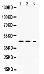
Anti-Neuroserpin Picoband Antibody
- SPECIFICATION
- CITATIONS
- PROTOCOLS
- BACKGROUND

Application
| WB |
|---|---|
| Primary Accession | Q99574 |
| Host | Rabbit |
| Reactivity | Human, Mouse, Rat |
| Clonality | Polyclonal |
| Format | Lyophilized |
| Description | Rabbit IgG polyclonal antibody for Neuroserpin(SERPINI1) detection. Tested with WB in Human;Mouse;Rat. |
| Reconstitution | Add 0.2ml of distilled water will yield a concentration of 500ug/ml. |
| Gene ID | 5274 |
|---|---|
| Other Names | Neuroserpin, Peptidase inhibitor 12, PI-12, Serpin I1, SERPINI1, PI12 |
| Calculated MW | 46427 MW KDa |
| Application Details | Western blot, 0.1-0.5 µg/ml, Human, Mouse, Rat |
| Subcellular Localization | Secreted. |
| Tissue Specificity | Predominantly expressed in the brain. |
| Protein Name | Neuroserpin |
| Contents | Each vial contains 5mg BSA, 0.9mg NaCl, 0.2mg Na2HPO4, 0.05mg NaN3. |
| Immunogen | A synthetic peptide corresponding to a sequence at the C-terminus of human Neuroserpin (272-310aa KAQLVEEWANSVKKQKVEVYLPRFTVEQEIDLKDVLKA L), different from the related mouse sequence by two amino acids, and from the related rat sequence by three amino aci |
| Purification | Immunogen affinity purified. |
| Cross Reactivity | No cross reactivity with other proteins. |
| Storage | At -20˚C for one year. After r˚Constitution, at 4˚C for one month. It˚Can also be aliquotted and stored frozen at -20˚C for a longer time.Avoid repeated freezing and thawing. |
| Name | SERPINI1 |
|---|---|
| Synonyms | PI12 |
| Function | Serine protease inhibitor that inhibits plasminogen activators and plasmin but not thrombin (PubMed:11880376, PubMed:19265707, PubMed:19285087, PubMed:26329378, PubMed:9442076). May be involved in the formation or reorganization of synaptic connections as well as for synaptic plasticity in the adult nervous system. May protect neurons from cell damage by tissue-type plasminogen activator (Probable). |
| Cellular Location | Secreted. Cytoplasmic vesicle, secretory vesicle lumen. Perikaryon |
| Tissue Location | Detected in brain cortex and hippocampus pyramidal neurons (at protein level) (PubMed:17040209). Detected in cerebrospinal fluid (at protein level) (PubMed:25326458). Predominantly expressed in the brain (PubMed:9070919). |

Thousands of laboratories across the world have published research that depended on the performance of antibodies from Abcepta to advance their research. Check out links to articles that cite our products in major peer-reviewed journals, organized by research category.
info@abcepta.com, and receive a free "I Love Antibodies" mug.

Provided below are standard protocols that you may find useful for product applications.
Background
Neuroserpin is a protein that in humans is encoded by the SERPINI1 gene. This gene encodes a member of the serpin superfamily of serine proteinase inhibitors. The protein is primarily secreted by axons in the brain, and preferentially reacts with and inhibits tissue-type plasminogen activator. It is thought to play a role in the regulation of axonal growth and the development of synaptic plasticity. Mutations in this gene result in familial encephalopathy with neuroserpin inclusion bodies (FENIB), which is a dominantly inherited form of familial encephalopathy and epilepsy characterized by the accumulation of mutant neuroserpin polymers. Multiple alternatively spliced variants, encoding the same protein, have been identified.
If you have used an Abcepta product and would like to share how it has performed, please click on the "Submit Review" button and provide the requested information. Our staff will examine and post your review and contact you if needed.
If you have any additional inquiries please email technical services at tech@abcepta.com.
Ordering Information
Other Products
Shipping Information
