


 Foundational characteristics of cancer include proliferation, angiogenesis, migration, evasion of apoptosis, and cellular immortality. Find key markers for these cellular processes and antibodies to detect them.
 The SUMOplot™ Analysis Program predicts and scores sumoylation sites in your protein. SUMOylation is a post-translational modification involved in various cellular processes, such as nuclear-cytosolic transport, transcriptional regulation, apoptosis, protein stability, response to stress, and progression through the cell cycle.
 The Autophagy Receptor Motif Plotter predicts and scores autophagy receptor binding sites in your protein. Identifying proteins connected to this pathway is critical to understanding the role of autophagy in physiological as well as pathological processes such as development, differentiation, neurodegenerative diseases, stress, infection, and cancer.
Anti-Lck Picoband Antibody
- SPECIFICATION
- CITATIONS
- PROTOCOLS
- BACKGROUND

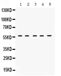
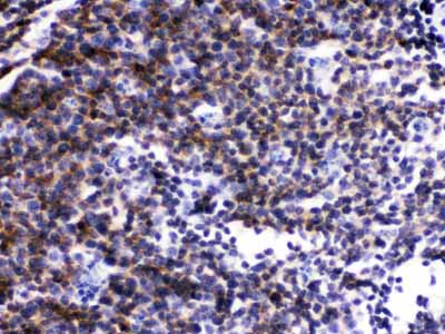
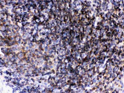
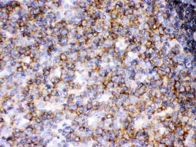
Application
| WB, IHC-P |
|---|---|
| Primary Accession | P06239 |
| Host | Rabbit |
| Reactivity | Human, Mouse, Rat |
| Clonality | Polyclonal |
| Format | Lyophilized |
| Description | Rabbit IgG polyclonal antibody for Tyrosine-protein kinase Lck(LCK) detection. Tested with WB, IHC-P in Human;Mouse;Rat. |
| Reconstitution | Add 0.2ml of distilled water will yield a concentration of 500ug/ml. |
| Gene ID | 3932 |
|---|---|
| Other Names | Tyrosine-protein kinase Lck, 2.7.10.2, Leukocyte C-terminal Src kinase, LSK, Lymphocyte cell-specific protein-tyrosine kinase, Protein YT16, Proto-oncogene Lck, T cell-specific protein-tyrosine kinase, p56-LCK, LCK |
| Calculated MW | 58001 MW KDa |
| Application Details | Immunohistochemistry(Paraffin-embedded Section), 0.5-1 µg/ml, Human, Mouse, Rat, By Heat Western blot, 0.1-0.5 µg/ml, Human |
| Subcellular Localization | Cytoplasm . Cell membrane ; Lipid-anchor ; Cytoplasmic side . Present in lipid rafts in an inactive form. |
| Tissue Specificity | Expressed specifically in lymphoid cells. |
| Protein Name | Tyrosine-protein kinase Lck |
| Contents | Each vial contains 5mg BSA, 0.9mg NaCl, 0.2mg Na2HPO4, 0.05mg NaN3. |
| Immunogen | A synthetic peptide corresponding to a sequence at the C-terminus of human Lck (468-506aa ELYQLMRLCWKERPEDRPTFDYLRSVLEDFFTATEGQYQ), different from the related mouse and rat sequences by three amino acids. |
| Purification | Immunogen affinity purified. |
| Cross Reactivity | No cross reactivity with other proteins. |
| Storage | At -20˚C for one year. After r˚Constitution, at 4˚C for one month. It˚Can also be aliquotted and stored frozen at -20˚C for a longer time.Avoid repeated freezing and thawing. |
| Name | LCK |
|---|---|
| Function | Non-receptor tyrosine-protein kinase that plays an essential role in the selection and maturation of developing T-cells in the thymus and in the function of mature T-cells (PubMed:2470098). Plays a key role in T-cell antigen receptor (TCR)-linked signal transduction pathways (PubMed:2470098). Constitutively associated with the cytoplasmic portions of the CD4 and CD8 surface receptors (PubMed:2470098). Association of the TCR with a peptide antigen-bound MHC complex facilitates the interaction of CD4 and CD8 with MHC class II and class I molecules, respectively, thereby recruiting the associated LCK protein to the vicinity of the TCR-CD3 complex (PubMed:2470098). LCK then phosphorylates tyrosine residues within the immunoreceptor tyrosine-based activation motifs (ITAM) of the cytoplasmic tails of the TCR-gamma chains and CD3 subunits, initiating the TCR-CD3 signaling pathway (PubMed:2470098, PubMed:40592325). Once stimulated, the TCR recruits the tyrosine kinase ZAP70, that becomes phosphorylated and activated by LCK. Following this, a large number of signaling molecules are recruited, ultimately leading to lymphokine production. LCK also contributes to signaling by other receptor molecules. Associates directly with the cytoplasmic tail of CD2, which leads to hyperphosphorylation and activation of LCK. Also plays a role in the IL2 receptor-linked signaling pathway that controls the T-cell proliferative response. Binding of IL2 to its receptor results in increased activity of LCK. Is expressed at all stages of thymocyte development and is required for the regulation of maturation events that are governed by both pre-TCR and mature alpha beta TCR. Phosphorylates other substrates including RUNX3, PTK2B/PYK2, the microtubule-associated protein MAPT, RHOH or TYROBP. Interacts with FYB2 (PubMed:27335501). |
| Cellular Location | Cell membrane; Lipid-anchor; Cytoplasmic side Cytoplasm, cytosol. Note=Present in lipid rafts in an inactive form. |
| Tissue Location | Expressed specifically in lymphoid cells. |

Thousands of laboratories across the world have published research that depended on the performance of antibodies from Abcepta to advance their research. Check out links to articles that cite our products in major peer-reviewed journals, organized by research category.
info@abcepta.com, and receive a free "I Love Antibodies" mug.

Provided below are standard protocols that you may find useful for product applications.
Background
Lck (or lymphocyte-specific protein tyrosine kinase) is a 56 kDa protein that is found inside specializedcells of the immune system called lymphocytes. The human LCK gene is mapped to chromosome 1p35-p32. This gene is a member of the Src family of protein tyrosine kinases (PTKs). The encoded protein is a key signaling molecule in the selection and maturation of developing T-cells. It contains N-terminal sites for myristylation and palmitylation, a PTK domain, and SH2 and SH3 domains which are involved in mediating protein-protein interactions with phosphotyrosine-containing and proline-rich motifs, respectively. The protein localizes to the plasma membrane and pericentrosomal vesicles, and binds to cell surface receptors, including CD4 and CD8, and other signaling molecules. Multiple alternatively spliced variants, encoding the same protein, have been described.
If you have used an Abcepta product and would like to share how it has performed, please click on the "Submit Review" button and provide the requested information. Our staff will examine and post your review and contact you if needed.
If you have any additional inquiries please email technical services at tech@abcepta.com.
Ordering Information
Other Products
Shipping Information
