


 Foundational characteristics of cancer include proliferation, angiogenesis, migration, evasion of apoptosis, and cellular immortality. Find key markers for these cellular processes and antibodies to detect them.
 The SUMOplot™ Analysis Program predicts and scores sumoylation sites in your protein. SUMOylation is a post-translational modification involved in various cellular processes, such as nuclear-cytosolic transport, transcriptional regulation, apoptosis, protein stability, response to stress, and progression through the cell cycle.
 The Autophagy Receptor Motif Plotter predicts and scores autophagy receptor binding sites in your protein. Identifying proteins connected to this pathway is critical to understanding the role of autophagy in physiological as well as pathological processes such as development, differentiation, neurodegenerative diseases, stress, infection, and cancer.
Anti-TPP1 Picoband Antibody
- SPECIFICATION
- CITATIONS
- PROTOCOLS
- BACKGROUND

Application
| WB, IHC-P |
|---|---|
| Primary Accession | O14773 |
| Host | Rabbit |
| Reactivity | Human |
| Clonality | Polyclonal |
| Format | Lyophilized |
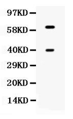
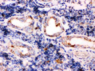
| Description | Rabbit IgG polyclonal antibody for Tripeptidyl-peptidase 1(TPP1) detection. Tested with WB, IHC-P in Human. |
| Reconstitution | Add 0.2ml of distilled water will yield a concentration of 500ug/ml. |
| Gene ID | 1200 |
|---|---|
| Other Names | Tripeptidyl-peptidase 1, TPP-1, 3.4.14.9, Cell growth-inhibiting gene 1 protein, Lysosomal pepstatin-insensitive protease, LPIC, Tripeptidyl aminopeptidase, Tripeptidyl-peptidase I, TPP-I, TPP1, CLN2 |
| Calculated MW | 61248 MW KDa |
| Application Details | Immunohistochemistry(Paraffin-embedded Section), 0.5-1 µg/ml, Human, By Heat Western blot, 0.1-0.5 µg/ml, Human |
| Subcellular Localization | Lysosome. Melanosome. Identified by mass spectrometry in melanosome fractions from stage I to stage IV. |
| Tissue Specificity | Detected in all tissues examined with highest levels in heart and placenta and relatively similar levels in other tissues. |
| Protein Name | Tripeptidyl-peptidase 1 |
| Contents | Each vial contains 5mg BSA, 0.9mg NaCl, 0.2mg Na2HPO4, 0.05mg NaN3. |
| Immunogen | A synthetic peptide corresponding to a sequence in the middle region of human TPP1 (227-261aa CAQFLEQYFHDSDLAQFMRLFGGNFAHQASVARVV), different from the related mouse sequence by six amino acids, and from the related rat sequence by five amino acids. |
| Purification | Immunogen affinity purified. |
| Cross Reactivity | No cross reactivity with other proteins. |
| Storage | At -20˚C for one year. After r˚Constitution, at 4˚C for one month. It˚Can also be aliquotted and stored frozen at -20˚C for a longer time.Avoid repeated freezing and thawing. |
| Name | TPP1 |
|---|---|
| Synonyms | CLN2 |
| Function | Lysosomal serine protease with tripeptidyl-peptidase I activity (PubMed:11054422, PubMed:19038966, PubMed:19038967). May act as a non-specific lysosomal peptidase which generates tripeptides from the breakdown products produced by lysosomal proteinases (PubMed:11054422, PubMed:19038966, PubMed:19038967). Requires substrates with an unsubstituted N-terminus (PubMed:19038966). |
| Cellular Location | Lysosome. Melanosome. Note=Identified by mass spectrometry in melanosome fractions from stage I to stage IV |
| Tissue Location | Detected in all tissues examined with highest levels in heart and placenta and relatively similar levels in other tissues |

Thousands of laboratories across the world have published research that depended on the performance of antibodies from Abcepta to advance their research. Check out links to articles that cite our products in major peer-reviewed journals, organized by research category.
info@abcepta.com, and receive a free "I Love Antibodies" mug.

Provided below are standard protocols that you may find useful for product applications.
Background
Tripeptidyl-peptidase 1, also known as Lysosomal pepstatin-insensitive protease, is an enzyme that in humans is encoded by the TPP1 gene. This gene encodes a member of the sedolisin family of serine proteases. The protease functions in the lysosome to cleave N-terminal tripeptides from substrates, and has weaker endopeptidase activity. It is synthesized as a catalytically-inactive enzyme which is activated and auto-proteolyzed upon acidification. Mutations in this gene result in late-infantile neuronal ceroid lipofuscinosis, which is associated with the failure to degrade specific neuropeptides and a subunit of ATP synthase in the lysosome.
If you have used an Abcepta product and would like to share how it has performed, please click on the "Submit Review" button and provide the requested information. Our staff will examine and post your review and contact you if needed.
If you have any additional inquiries please email technical services at tech@abcepta.com.
Ordering Information
Other Products
Shipping Information
