


 Foundational characteristics of cancer include proliferation, angiogenesis, migration, evasion of apoptosis, and cellular immortality. Find key markers for these cellular processes and antibodies to detect them.
 The SUMOplot™ Analysis Program predicts and scores sumoylation sites in your protein. SUMOylation is a post-translational modification involved in various cellular processes, such as nuclear-cytosolic transport, transcriptional regulation, apoptosis, protein stability, response to stress, and progression through the cell cycle.
 The Autophagy Receptor Motif Plotter predicts and scores autophagy receptor binding sites in your protein. Identifying proteins connected to this pathway is critical to understanding the role of autophagy in physiological as well as pathological processes such as development, differentiation, neurodegenerative diseases, stress, infection, and cancer.
Anti-CD30/TNFRSF8 Antibody
- SPECIFICATION
- CITATIONS
- PROTOCOLS
- BACKGROUND

Application
| WB |
|---|---|
| Primary Accession | P28908 |
| Host | Rabbit |
| Reactivity | Human |
| Clonality | Polyclonal |
| Format | Lyophilized |
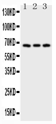
| Description | Rabbit IgG polyclonal antibody for Tumor necrosis factor receptor superfamily member 8(TNFRSF8) detection. Tested with WB in Human. |
| Reconstitution | Add 0.2ml of distilled water will yield a concentration of 500ug/ml. |
| Gene ID | 943 |
|---|---|
| Other Names | Tumor necrosis factor receptor superfamily member 8, CD30L receptor, Ki-1 antigen, Lymphocyte activation antigen CD30, CD30, TNFRSF8, CD30, D1S166E |
| Calculated MW | 63747 MW KDa |
| Application Details | Western blot, 0.1-0.5 µg/ml, Human |
| Subcellular Localization | Isoform 1: Cell membrane; Single-pass type I membrane protein. |
| Protein Name | Tumor necrosis factor receptor superfamily member 8 |
| Contents | Each vial contains 5mg BSA, 0.9mg NaCl, 0.2mg Na2HPO4, 0.05mg NaN3. |
| Immunogen | E.coli-derived human CD30 recombinant protein (Position: T322-K595). Human CD30 shares 60% and 57% amino acid (aa) sequences identity with mouse and rat CD30, respectively. |
| Purification | Immunogen affinity purified. |
| Cross Reactivity | No cross reactivity with other proteins |
| Storage | At -20˚C for one year. After r˚Constitution, at 4˚C for one month. It˚Can also be aliquotted and stored frozen at -20˚C for a longer time.Avoid repeated freezing and thawing. |
| Sequence Similarities | Contains 6 TNFR-Cys repeats. |
| Name | TNFRSF8 (HGNC:11923) |
|---|---|
| Function | Receptor for TNFSF8/CD30L (PubMed:8391931). May play a role in the regulation of cellular growth and transformation of activated lymphoblasts. Regulates gene expression through activation of NF-kappa- B (PubMed:8999898). |
| Cellular Location | [Isoform 1]: Cell membrane; Single-pass type I membrane protein |
| Tissue Location | [Isoform 2]: Detected in alveolar macrophages (at protein level). |

Thousands of laboratories across the world have published research that depended on the performance of antibodies from Abcepta to advance their research. Check out links to articles that cite our products in major peer-reviewed journals, organized by research category.
info@abcepta.com, and receive a free "I Love Antibodies" mug.

Provided below are standard protocols that you may find useful for product applications.
Background
CD30, also known as TNFRSF8, is a cell membrane protein of the tumor necrosis factor receptor family and tumor marker. This receptor is a positive regulator of apoptosis, and also has been shown to limit the proliferative potential of autoreactive CD8 effector T cells and protect the body against autoimmunity. This gene is mapped to 1p36.22. CD30 is expressed in embryonal carcinoma but not in seminoma and is thus a useful marker in distinguishing between these germ cell tumors. CD30 mast cell activation represents an IgE-independent activation pathway, which is important for understanding cutaneous inflammation associated with mast cells. In addition to those, CD30 is also associated with anaplastic large cell lymphoma.
If you have used an Abcepta product and would like to share how it has performed, please click on the "Submit Review" button and provide the requested information. Our staff will examine and post your review and contact you if needed.
If you have any additional inquiries please email technical services at tech@abcepta.com.
Ordering Information
Other Products
Shipping Information
