


 Foundational characteristics of cancer include proliferation, angiogenesis, migration, evasion of apoptosis, and cellular immortality. Find key markers for these cellular processes and antibodies to detect them.
 The SUMOplot™ Analysis Program predicts and scores sumoylation sites in your protein. SUMOylation is a post-translational modification involved in various cellular processes, such as nuclear-cytosolic transport, transcriptional regulation, apoptosis, protein stability, response to stress, and progression through the cell cycle.
 The Autophagy Receptor Motif Plotter predicts and scores autophagy receptor binding sites in your protein. Identifying proteins connected to this pathway is critical to understanding the role of autophagy in physiological as well as pathological processes such as development, differentiation, neurodegenerative diseases, stress, infection, and cancer.
Anti-PD-1 Antibody
- SPECIFICATION
- CITATIONS
- PROTOCOLS
- BACKGROUND

Application
| WB |
|---|---|
| Primary Accession | Q15116 |
| Host | Rabbit |
| Reactivity | Human, Rat |
| Clonality | Polyclonal |
| Format | Lyophilized |
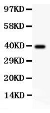
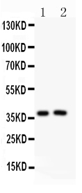
| Description | Rabbit IgG polyclonal antibody for Programmed cell death protein 1(PDCD1) detection. Tested with WB in Human;Rat. |
| Reconstitution | Add 0.2ml of distilled water will yield a concentration of 500ug/ml. |
| Gene ID | 5133 |
|---|---|
| Other Names | Programmed cell death protein 1, Protein PD-1, hPD-1, CD279, PDCD1, PD1 |
| Calculated MW | 31647 MW KDa |
| Application Details | Western blot, 0.1-0.5 µg/ml, Human, Rat |
| Subcellular Localization | Membrane; Single-pass type I membrane protein. |
| Protein Name | Programmed cell death protein 1 |
| Contents | Each vial contains 5mg BSA, 0.9mg NaCl, 0.2mg Na2HPO4, 0.05mg NaN3. |
| Immunogen | E.coli-derived human PD1 recombinant protein (Position: P101-L288). Human PD1 shares 59% amino acid (aa) sequence identity with mouse PD1. |
| Purification | Immunogen affinity purified. |
| Cross Reactivity | No cross reactivity with other proteins |
| Storage | At -20˚C for one year. After r˚Constitution, at 4˚C for one month. It˚Can also be aliquotted and stored frozen at -20˚C for a longer time.Avoid repeated freezing and thawing. |
| Name | PDCD1 {ECO:0000303|PubMed:7851902, ECO:0000312|HGNC:HGNC:8760} |
|---|---|
| Function | Inhibitory receptor on antigen activated T-cells that plays a critical role in induction and maintenance of immune tolerance to self (PubMed:21276005, PubMed:37208329). Delivers inhibitory signals upon binding to ligands CD274/PDCD1L1 and CD273/PDCD1LG2 (PubMed:21276005). Following T-cell receptor (TCR) engagement, PDCD1 associates with CD3- TCR in the immunological synapse and directly inhibits T-cell activation (By similarity). Suppresses T-cell activation through the recruitment of PTPN11/SHP-2: following ligand-binding, PDCD1 is phosphorylated within the ITSM motif, leading to the recruitment of the protein tyrosine phosphatase PTPN11/SHP-2 that mediates dephosphorylation of key TCR proximal signaling molecules, such as ZAP70, PRKCQ/PKCtheta and CD247/CD3zeta (By similarity). |
| Cellular Location | Cell membrane; Single-pass type I membrane protein |

Thousands of laboratories across the world have published research that depended on the performance of antibodies from Abcepta to advance their research. Check out links to articles that cite our products in major peer-reviewed journals, organized by research category.
info@abcepta.com, and receive a free "I Love Antibodies" mug.

Provided below are standard protocols that you may find useful for product applications.
Background
PDCD1(Programmed cell death 1), also called PD1, encodes a cell surface receptor that is a member of the B7 superfamily involved in immunomodulation. This gene is mapped to 2q37.3. PDCD1 acts as an inhibitory molecule on T cells after interacting with its ligands PDL1 and PDL2. The PDCD1 gene contains 5 exons. This protein is expressed in pro-B-cells and is thought to play a role in their differentiation. Using flow cytometric analysis, It has been found that expression of PDCD1 was upregulated on CD16-positive and CD16-negative monocytes, but not on dendritic cells, in viremic HIV-positive patients, but not in highly active antiretroviral therapy (HAART)-treated HIV-positive patients. PDCD1 upregulation in monocytes was induced by microbial Toll-like receptor ligands and inflammatory cytokines.
If you have used an Abcepta product and would like to share how it has performed, please click on the "Submit Review" button and provide the requested information. Our staff will examine and post your review and contact you if needed.
If you have any additional inquiries please email technical services at tech@abcepta.com.
Ordering Information
Other Products
Shipping Information
