


 Foundational characteristics of cancer include proliferation, angiogenesis, migration, evasion of apoptosis, and cellular immortality. Find key markers for these cellular processes and antibodies to detect them.
 The SUMOplot™ Analysis Program predicts and scores sumoylation sites in your protein. SUMOylation is a post-translational modification involved in various cellular processes, such as nuclear-cytosolic transport, transcriptional regulation, apoptosis, protein stability, response to stress, and progression through the cell cycle.
 The Autophagy Receptor Motif Plotter predicts and scores autophagy receptor binding sites in your protein. Identifying proteins connected to this pathway is critical to understanding the role of autophagy in physiological as well as pathological processes such as development, differentiation, neurodegenerative diseases, stress, infection, and cancer.
Anti-PKC Delta Antibody
- SPECIFICATION
- CITATIONS
- PROTOCOLS
- BACKGROUND

Application
| WB |
|---|---|
| Primary Accession | Q05655 |
| Host | Rabbit |
| Reactivity | Human |
| Clonality | Polyclonal |
| Format | Lyophilized |
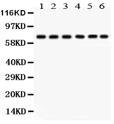
| Description | Rabbit IgG polyclonal antibody for Protein kinase C delta type(PRKCD) detection. Tested with WB in Human. |
| Reconstitution | Add 0.2ml of distilled water will yield a concentration of 500ug/ml. |
| Gene ID | 5580 |
|---|---|
| Other Names | Protein kinase C delta type, 2.7.11.13, Tyrosine-protein kinase PRKCD, 2.7.10.2, nPKC-delta, Protein kinase C delta type regulatory subunit, Protein kinase C delta type catalytic subunit, Sphingosine-dependent protein kinase-1, SDK1, PRKCD |
| Calculated MW | 77505 MW KDa |
| Application Details | Western blot, 0.1-0.5 µg/ml, Human |
| Subcellular Localization | Cytoplasm. Cytoplasm, perinuclear region. Nucleus. Endoplasmic reticulum. Mitochondrion. Cell membrane; Peripheral membrane protein. |
| Protein Name | Protein kinase C delta type |
| Contents | Each vial contains 5mg BSA, 0.9mg NaCl, 0.2mg Na2HPO4, 0.05mg NaN3. |
| Immunogen | E.coli-derived human PKC delta recombinant protein (Position: M1-E160). Human PKC delta shares 95% and 93% amino acid (aa) sequence identity with mouse and rat PKC delta, respctively. |
| Purification | Immunogen affinity purified. |
| Cross Reactivity | No cross reactivity with other proteins |
| Storage | At -20˚C for one year. After r˚Constitution, at 4˚C for one month. It˚Can also be aliquotted and stored frozen at -20˚C for a longer time.Avoid repeated freezing and thawing. |
| Sequence Similarities | Belongs to the protein kinase superfamily. AGC Ser/Thr protein kinase family. PKC subfamily. |
| Name | PRKCD (HGNC:9399) |
|---|---|
| Function | Calcium-independent, phospholipid- and diacylglycerol (DAG)- dependent serine/threonine-protein kinase that plays contrasting roles in cell death and cell survival by functioning as a pro-apoptotic protein during DNA damage-induced apoptosis, but acting as an anti- apoptotic protein during cytokine receptor-initiated cell death, is involved in tumor suppression as well as survival of several cancers, is required for oxygen radical production by NADPH oxidase and acts as positive or negative regulator in platelet functional responses (PubMed:21406692, PubMed:21810427). Negatively regulates B cell proliferation and also has an important function in self-antigen induced B cell tolerance induction (By similarity). Upon DNA damage, activates the promoter of the death-promoting transcription factor BCLAF1/Btf to trigger BCLAF1-mediated p53/TP53 gene transcription and apoptosis (PubMed:21406692, PubMed:21810427). In response to oxidative stress, interact with and activate CHUK/IKKA in the nucleus, causing the phosphorylation of p53/TP53 (PubMed:21406692, PubMed:21810427). In the case of ER stress or DNA damage-induced apoptosis, can form a complex with the tyrosine-protein kinase ABL1 which trigger apoptosis independently of p53/TP53 (PubMed:21406692, PubMed:21810427). In cytosol can trigger apoptosis by activating MAPK11 or MAPK14, inhibiting AKT1 and decreasing the level of X-linked inhibitor of apoptosis protein (XIAP), whereas in nucleus induces apoptosis via the activation of MAPK8 or MAPK9. Upon ionizing radiation treatment, is required for the activation of the apoptosis regulators BAX and BAK, which trigger the mitochondrial cell death pathway. Can phosphorylate MCL1 and target it for degradation which is sufficient to trigger for BAX activation and apoptosis. Is required for the control of cell cycle progression both at G1/S and G2/M phases. Mediates phorbol 12-myristate 13-acetate (PMA)-induced inhibition of cell cycle progression at G1/S phase by up-regulating the CDK inhibitor CDKN1A/p21 and inhibiting the cyclin CCNA2 promoter activity. In response to UV irradiation can phosphorylate CDK1, which is important for the G2/M DNA damage checkpoint activation (By similarity). Can protect glioma cells from the apoptosis induced by TNFSF10/TRAIL, probably by inducing increased phosphorylation and subsequent activation of AKT1 (PubMed:15774464). Is highly expressed in a number of cancer cells and promotes cell survival and resistance against chemotherapeutic drugs by inducing cyclin D1 (CCND1) and hyperphosphorylation of RB1, and via several pro-survival pathways, including NF-kappa-B, AKT1 and MAPK1/3 (ERK1/2). Involved in antifungal immunity by mediating phosphorylation and activation of CARD9 downstream of C-type lectin receptors activation, promoting interaction between CARD9 and BCL10, followed by activation of NF- kappa-B and MAP kinase p38 pathways (By similarity). Can also act as tumor suppressor upon mitogenic stimulation with PMA or TPA. In N- formyl-methionyl-leucyl-phenylalanine (fMLP)-treated cells, is required for NCF1 (p47-phox) phosphorylation and activation of NADPH oxidase activity, and regulates TNF-elicited superoxide anion production in neutrophils, by direct phosphorylation and activation of NCF1 or indirectly through MAPK1/3 (ERK1/2) signaling pathways (PubMed:19801500). May also play a role in the regulation of NADPH oxidase activity in eosinophil after stimulation with IL5, leukotriene B4 or PMA (PubMed:11748588). In collagen-induced platelet aggregation, acts a negative regulator of filopodia formation and actin polymerization by interacting with and negatively regulating VASP phosphorylation (PubMed:16940418). Downstream of PAR1, PAR4 and CD36/GP4 receptors, regulates differentially platelet dense granule secretion; acts as a positive regulator in PAR-mediated granule secretion, whereas it negatively regulates CD36/GP4-mediated granule release (PubMed:19587372). Phosphorylates MUC1 in the C-terminal and regulates the interaction between MUC1 and beta-catenin (PubMed:11877440). The catalytic subunit phosphorylates 14-3-3 proteins (YWHAB, YWHAZ and YWHAH) in a sphingosine-dependent fashion (By similarity). Phosphorylates ELAVL1 in response to angiotensin-2 treatment (PubMed:18285462). Phosphorylates mitochondrial phospholipid scramblase 3 (PLSCR3), resulting in increased cardiolipin expression on the mitochondrial outer membrane which facilitates apoptosis (PubMed:12649167). Phosphorylates SMPD1 which induces SMPD1 secretion (PubMed:17303575). |
| Cellular Location | Cytoplasm. Cytoplasm, perinuclear region. Nucleus. Cell membrane; Peripheral membrane protein Mitochondrion. Endomembrane system. Note=Translocates to the mitochondria upon apoptotic stimulation. Upon activation, translocates to the plasma membrane followed by partial location to the endolysosomes (PubMed:17303575). |

Thousands of laboratories across the world have published research that depended on the performance of antibodies from Abcepta to advance their research. Check out links to articles that cite our products in major peer-reviewed journals, organized by research category.
info@abcepta.com, and receive a free "I Love Antibodies" mug.

Provided below are standard protocols that you may find useful for product applications.
Background
Protein kinase C delta type, also called PKCD, is an enzyme that in humans is encoded by the PRKCD gene. The PRKCD gene encodes a member of the protein kinase C family, members of which are critical for regulation of cell survival, proliferation, and apoptosis. This gene is mapped to 3p21.1. Studies both in human and mice demonstrate that this kinase is involved inB cell signaling and in the regulation of growth, apoptosis, and differentiation of a variety of cell types. PRKCD is also regulated by phosphorylation on various tyrosine residues including Y311 (by SRC). It has been showed that PRKCD phosphorylates NLRC4 and that this phosphorylation is critical for inflammasome assembly. What’s more, it is also a part of a noncanonical WNT signaling cascade.
If you have used an Abcepta product and would like to share how it has performed, please click on the "Submit Review" button and provide the requested information. Our staff will examine and post your review and contact you if needed.
If you have any additional inquiries please email technical services at tech@abcepta.com.
Ordering Information
Other Products
Shipping Information
