


 Foundational characteristics of cancer include proliferation, angiogenesis, migration, evasion of apoptosis, and cellular immortality. Find key markers for these cellular processes and antibodies to detect them.
 The SUMOplot™ Analysis Program predicts and scores sumoylation sites in your protein. SUMOylation is a post-translational modification involved in various cellular processes, such as nuclear-cytosolic transport, transcriptional regulation, apoptosis, protein stability, response to stress, and progression through the cell cycle.
 The Autophagy Receptor Motif Plotter predicts and scores autophagy receptor binding sites in your protein. Identifying proteins connected to this pathway is critical to understanding the role of autophagy in physiological as well as pathological processes such as development, differentiation, neurodegenerative diseases, stress, infection, and cancer.
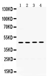
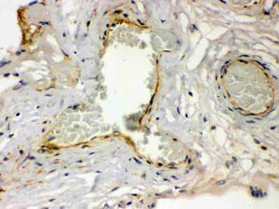
Anti-Thrombin Receptor Antibody
- SPECIFICATION
- CITATIONS
- PROTOCOLS
- BACKGROUND

Application
| WB, IHC-P |
|---|---|
| Primary Accession | P25116 |
| Host | Rabbit |
| Reactivity | Human |
| Clonality | Polyclonal |
| Format | Lyophilized |
| Description | Rabbit IgG polyclonal antibody for Proteinase-activated receptor 1(F2R) detection. Tested with WB, IHC-P in Human. |
| Reconstitution | Add 0.2ml of distilled water will yield a concentration of 500ug/ml. |
| Gene ID | 2149 |
|---|---|
| Other Names | Proteinase-activated receptor 1, PAR-1, Coagulation factor II receptor, Thrombin receptor, F2R, CF2R, PAR1, TR |
| Calculated MW | 47441 MW KDa |
| Application Details | Immunohistochemistry(Paraffin-embedded Section), 0.5-1 µg/ml, Human, By Heat Western blot, 0.1-0.5 µg/ml, Human |
| Subcellular Localization | Cell membrane; Multi-pass membrane protein. |
| Tissue Specificity | Platelets and vascular endothelial cells. |
| Protein Name | Proteinase-activated receptor 1 |
| Contents | Each vial contains 5mg BSA, 0.9mg NaCl, 0.2mg Na2HPO4, 0.05mg NaN3. |
| Immunogen | A synthetic peptide corresponding to a sequence at the N-terminus of human Thrombin Receptor (46-82aa RNPNDKYEPFWEDEEKNESGLTEYRLVSINKSSPLQK). |
| Purification | Immunogen affinity purified. |
| Cross Reactivity | No cross reactivity with other proteins |
| Storage | At -20˚C for one year. After r˚Constitution, at 4˚C for one month. It˚Can also be aliquotted and stored frozen at -20˚C for a longer time.Avoid repeated freezing and thawing. |
| Name | F2R (HGNC:3537) |
|---|---|
| Synonyms | CF2R, PAR1, TR |
| Function | High affinity receptor that binds the activated thrombin, leading to calcium release from intracellular stores (PubMed:1672265, PubMed:8136362). The thrombin-activated receptor signaling pathway is mediated through PTX-insensitive G proteins, activation of phospholipase C resulting in the production of 1D-myo-inositol 1,4,5- trisphosphate (InsP3) which binds to InsP3 receptors causing calcium release from the stores (By similarity). In astrocytes, the calcium released into the cytosol allows the Ca(2+)-dependent release of L- glutamate into the synaptic cleft through BEST1, that targets the neuronal postsynaptic GRIN2A/NMDAR receptor resulting in the synaptic plasticity regulation (By similarity). May play a role in platelets activation and in vascular development (PubMed:10079109). Mediates up- regulation of pro-inflammatory cytokines, such as MCP-1/CCL2 and IL6, triggered by coagulation factor Xa (F10) in cardiac fibroblasts and umbilical vein endothelial cells (PubMed:30568593, PubMed:34831181). |
| Cellular Location | Cell membrane {ECO:0000250|UniProtKB:P26824}; Multi-pass membrane protein {ECO:0000250|UniProtKB:P26824} |
| Tissue Location | Platelets and vascular endothelial cells. |

Thousands of laboratories across the world have published research that depended on the performance of antibodies from Abcepta to advance their research. Check out links to articles that cite our products in major peer-reviewed journals, organized by research category.
info@abcepta.com, and receive a free "I Love Antibodies" mug.

Provided below are standard protocols that you may find useful for product applications.
Background
Proteinase-activated receptor 1 (PAR1), also known as the coagulation factor II (thrombin) receptor, is a protein that in humans is encoded by the F2R gene. By fluorescence in situ hybridization, this gene is mapped to 5q13, confirming its presence as a single locus in the human genome. PAR1 is a G protein-coupled receptor involved in the regulation of thrombotic response. Proteolytic cleavage leads to the activation of the receptor. The expression of PAR1 is both required and sufficient to promote growth and invasion of breast carcinoma cells in a xenograft mouse model.Â
If you have used an Abcepta product and would like to share how it has performed, please click on the "Submit Review" button and provide the requested information. Our staff will examine and post your review and contact you if needed.
If you have any additional inquiries please email technical services at tech@abcepta.com.
Ordering Information
Other Products
Shipping Information
