


 Foundational characteristics of cancer include proliferation, angiogenesis, migration, evasion of apoptosis, and cellular immortality. Find key markers for these cellular processes and antibodies to detect them.
 The SUMOplot™ Analysis Program predicts and scores sumoylation sites in your protein. SUMOylation is a post-translational modification involved in various cellular processes, such as nuclear-cytosolic transport, transcriptional regulation, apoptosis, protein stability, response to stress, and progression through the cell cycle.
 The Autophagy Receptor Motif Plotter predicts and scores autophagy receptor binding sites in your protein. Identifying proteins connected to this pathway is critical to understanding the role of autophagy in physiological as well as pathological processes such as development, differentiation, neurodegenerative diseases, stress, infection, and cancer.
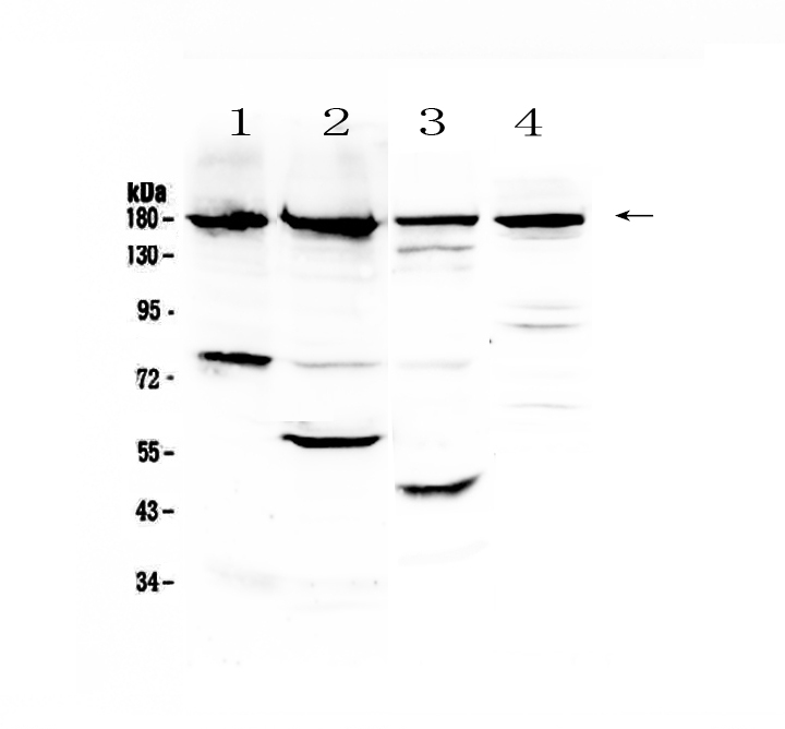
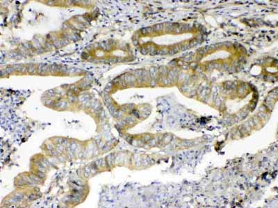
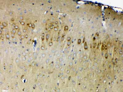
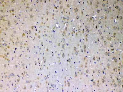
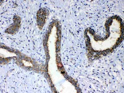
Anti-ErbB 4 Picoband Antibody
- SPECIFICATION
- CITATIONS
- PROTOCOLS
- BACKGROUND

Application
| WB, IHC-P |
|---|---|
| Primary Accession | Q15303 |
| Host | Rabbit |
| Reactivity | Human, Mouse, Rat |
| Clonality | Polyclonal |
| Format | Lyophilized |
| Description | Rabbit IgG polyclonal antibody for ErbB 4 detection. Tested with WB, IHC-P in Human;Mouse;Rat. |
| Reconstitution | Add 0.2ml of distilled water will yield a concentration of 500ug/ml. |
| Gene ID | 2066 |
|---|---|
| Other Names | Receptor tyrosine-protein kinase erbB-4, 2.7.10.1, Proto-oncogene-like protein c-ErbB-4, Tyrosine kinase-type cell surface receptor HER4, p180erbB4, ERBB4 intracellular domain, 4ICD, E4ICD, s80HER4, ERBB4, HER4 |
| Calculated MW | 146808 Da |
| Application Details | Western blot, 0.1-0.5 µg/ml Immunohistochemistry(Paraffin-embedded Section), 0.5-1 µg/ml |
| Subcellular Localization | Cell membrane. |
| Tissue Specificity | Expressed at highest levels in brain, heart, kidney, in addition to skeletal muscle, parathyroid, cerebellum, pituitary, spleen, testis and breast. Lower levels in thymus, lung, salivary gland, and pancreas. Isoform JM-A CYT-1 and isoform JM-B CYT-1 are expressed in cerebellum, but only the isoform JM-B is expressed in the heart. |
| Contents | Each vial contains 4mg Trehalose, 0.9mg NaCl, 0.2mg Na2HPO4, 0.05mg NaN3. |
| Immunogen | A synthetic peptide corresponding to a sequence of human ErbB 4 (SLSDLEQQYRALRKYYENCEVVMGNLEITSIEHNRDLSFLR). |
| Cross Reactivity | No cross reactivity with other proteins. |
| Storage | At -20˚C; for one year. After r˚Constitution, at 4˚C; for one month. It˚Can also be aliquotted and stored frozen at -20˚C; for a longer time. Avoid repeated freezing and thawing. |
| Name | ERBB4 |
|---|---|
| Synonyms | HER4 |
| Function | Tyrosine-protein kinase that plays an essential role as cell surface receptor for neuregulins and EGF family members and regulates development of the heart, the central nervous system and the mammary gland, gene transcription, cell proliferation, differentiation, migration and apoptosis. Required for normal cardiac muscle differentiation during embryonic development, and for postnatal cardiomyocyte proliferation. Required for normal development of the embryonic central nervous system, especially for normal neural crest cell migration and normal axon guidance. Required for mammary gland differentiation, induction of milk proteins and lactation. Acts as cell-surface receptor for the neuregulins NRG1, NRG2, NRG3 and NRG4 and the EGF family members BTC, EREG and HBEGF. Ligand binding triggers receptor dimerization and autophosphorylation at specific tyrosine residues that then serve as binding sites for scaffold proteins and effectors. Ligand specificity and signaling is modulated by alternative splicing, proteolytic processing, and by the formation of heterodimers with other ERBB family members, thereby creating multiple combinations of intracellular phosphotyrosines that trigger ligand- and context- specific cellular responses. Mediates phosphorylation of SHC1 and activation of the MAP kinases MAPK1/ERK2 and MAPK3/ERK1. Isoform JM-A CYT-1 and isoform JM-B CYT-1 phosphorylate PIK3R1, leading to the activation of phosphatidylinositol 3-kinase and AKT1 and protect cells against apoptosis. Isoform JM-A CYT-1 and isoform JM-B CYT-1 mediate reorganization of the actin cytoskeleton and promote cell migration in response to NRG1. Isoform JM-A CYT-2 and isoform JM-B CYT-2 lack the phosphotyrosine that mediates interaction with PIK3R1, and hence do not phosphorylate PIK3R1, do not protect cells against apoptosis, and do not promote reorganization of the actin cytoskeleton and cell migration. Proteolytic processing of isoform JM-A CYT-1 and isoform JM- A CYT-2 gives rise to the corresponding soluble intracellular domains (4ICD) that translocate to the nucleus, promote nuclear import of STAT5A, activation of STAT5A, mammary epithelium differentiation, cell proliferation and activation of gene expression. The ERBB4 soluble intracellular domains (4ICD) colocalize with STAT5A at the CSN2 promoter to regulate transcription of milk proteins during lactation. The ERBB4 soluble intracellular domains can also translocate to mitochondria and promote apoptosis. |
| Cellular Location | Cell membrane; Single-pass type I membrane protein. Note=In response to NRG1 treatment, the activated receptor is internalized |
| Tissue Location | Expressed at highest levels in brain, heart, kidney, in addition to skeletal muscle, parathyroid, cerebellum, pituitary, spleen, testis and breast. Lower levels in thymus, lung, salivary gland, and pancreas. Isoform JM-A CYT-1 and isoform JM-B CYT-1 are expressed in cerebellum, but only the isoform JM-B is expressed in the heart. |

Thousands of laboratories across the world have published research that depended on the performance of antibodies from Abcepta to advance their research. Check out links to articles that cite our products in major peer-reviewed journals, organized by research category.
info@abcepta.com, and receive a free "I Love Antibodies" mug.

Provided below are standard protocols that you may find useful for product applications.
Background
ERBB4(V-erb-b2 avian erythroblastic leukemia viral oncogene homolog 4) also known as ONCOGENE ERBB4 or HER4, is an enzyme that in humans is encoded by the ERBB4 gene. The HER4/ERBB4 gene is a member of the type I receptor tyrosine kinase subfamily that includes EGFR, ERBB2 and ERBB3. This gene is mapped on 2q34. ERBB4 is a single-pass type I transmembrane protein with multiple furin-like cysteine rich domains, a tyrosine kinase domain, a phosphotidylinositol-3 kinase binding site and a PDZ domainbinding motif. Furthermore, ERBB4 is a transmembrane receptor tyrosine kinase that regulates cell proliferation and differentiation. After binding its ligand, heregulin, or activation of protein kinase C by TPA, the ERBB4 ectodomain is cleaved by a metalloprotease.
If you have used an Abcepta product and would like to share how it has performed, please click on the "Submit Review" button and provide the requested information. Our staff will examine and post your review and contact you if needed.
If you have any additional inquiries please email technical services at tech@abcepta.com.

Ordering Information
Other Products
Shipping Information
