


 Foundational characteristics of cancer include proliferation, angiogenesis, migration, evasion of apoptosis, and cellular immortality. Find key markers for these cellular processes and antibodies to detect them.
 The SUMOplot™ Analysis Program predicts and scores sumoylation sites in your protein. SUMOylation is a post-translational modification involved in various cellular processes, such as nuclear-cytosolic transport, transcriptional regulation, apoptosis, protein stability, response to stress, and progression through the cell cycle.
 The Autophagy Receptor Motif Plotter predicts and scores autophagy receptor binding sites in your protein. Identifying proteins connected to this pathway is critical to understanding the role of autophagy in physiological as well as pathological processes such as development, differentiation, neurodegenerative diseases, stress, infection, and cancer.
Anti-XRCC1 Picoband Antibody
- SPECIFICATION
- CITATIONS
- PROTOCOLS
- BACKGROUND

Application
| WB, IHC-P, E |
|---|---|
| Primary Accession | P18887 |
| Host | Rabbit |
| Reactivity | Human, Mouse, Rat |
| Clonality | Polyclonal |
| Format | Lyophilized |
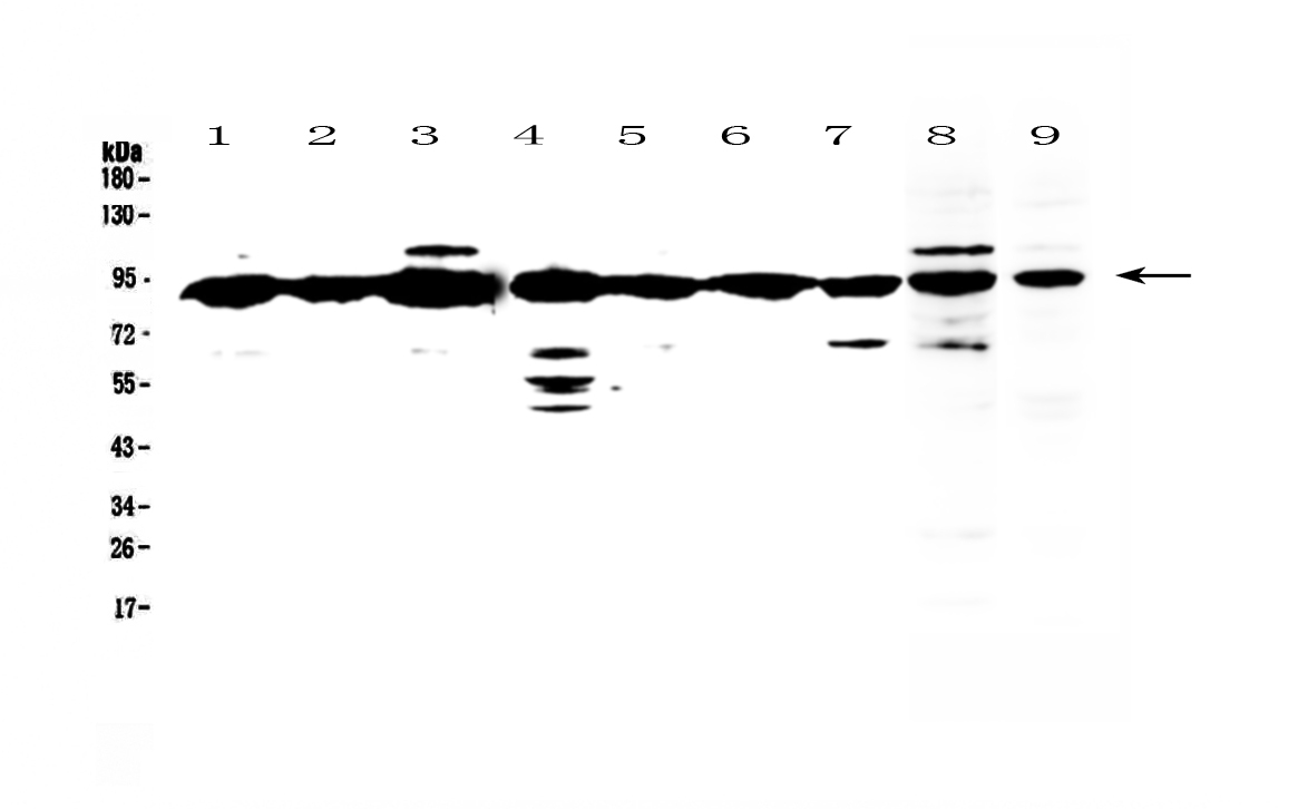
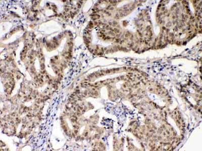
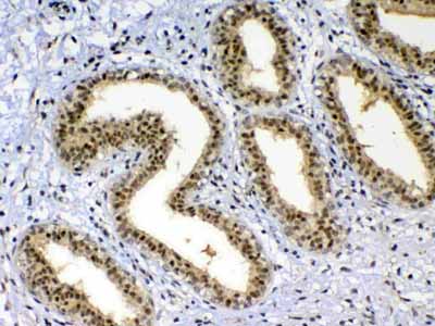
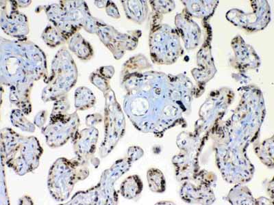
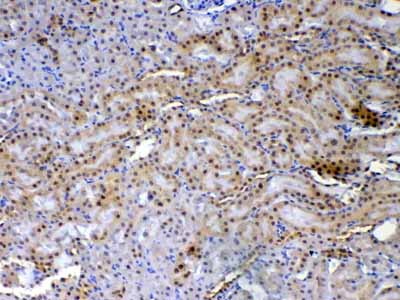
| Description | Rabbit IgG polyclonal antibody for XRCC1 detection. Tested with WB, IHC-P, Direct ELISA in Human;Mouse;Rat. |
| Reconstitution | Add 0.2ml of distilled water will yield a concentration of 500ug/ml. |
| Gene ID | 7515 |
|---|---|
| Other Names | DNA repair protein XRCC1, X-ray repair cross-complementing protein 1, XRCC1 |
| Calculated MW | 69498 Da |
| Application Details | Western blot, 0.1-0.5 µg/ml Immunohistochemistry(Paraffin-embedded Section), 0.5-1 µg/ml Direct ELISA, 0.1-0.5 µg/ml |
| Subcellular Localization | Nucleus. |
| Contents | Each vial contains 4mg Trehalose, 0.9mg NaCl, 0.2mg Na2HPO4, 0.05mg NaN3. |
| Immunogen | E. coli-derived human XRCC1 recombinant protein (Position: E538-A633). |
| Cross Reactivity | No cross reactivity with other proteins. |
| Storage | At -20˚C; for one year. After r˚Constitution, at 4˚C; for one month. It˚Can also be aliquotted and stored frozen at -20˚C; for a longer time. Avoid repeated freezing and thawing. |
| Name | XRCC1 {ECO:0000303|PubMed:2247054, ECO:0000312|HGNC:HGNC:12828} |
|---|---|
| Function | Scaffold protein involved in DNA single-strand break repair by mediating the assembly of DNA break repair protein complexes (PubMed:11163244, PubMed:28002403). Negatively regulates ADP- ribosyltransferase activity of PARP1 during base-excision repair in order to prevent excessive PARP1 activity (PubMed:28002403, PubMed:34102106, PubMed:34811483). Recognizes and binds poly-ADP-ribose chains: specifically binds auto-poly-ADP-ribosylated PARP1, limiting its activity (PubMed:14500814, PubMed:34102106, PubMed:34811483). |
| Cellular Location | Nucleus. Chromosome Note=Moves from the nucleoli to the global nuclear chromatin upon DNA damage (PubMed:28002403). Recruited to DNA damage sites fowwing interaction with poly-ADP-ribose chains (PubMed:14500814) |
| Tissue Location | Expressed in fibroblasts, retinal pigmented epithelial cells and lymphoblastoid cells (at protein level) |

Thousands of laboratories across the world have published research that depended on the performance of antibodies from Abcepta to advance their research. Check out links to articles that cite our products in major peer-reviewed journals, organized by research category.
info@abcepta.com, and receive a free "I Love Antibodies" mug.

Provided below are standard protocols that you may find useful for product applications.
Background
XRCC1(X-RAY REPAIR, COMPLEMENTING DEFECTIVE, IN CHINESE HAMSTER, 1) is a DNA repair protein which complexes with DNA ligase III. The protein encoded by this gene is involved in the efficient repair of DNA single-strand breaks formed by exposure to ionizing radiation and alkylating agents. The XRCC1 gene is mapped to 19q13.31. The XRCC1 interacts with DNA ligase III, polymerase beta and poly (ADP-ribose) polymerase to participate in the base excision repair pathway. It may play a role in DNA processing during meiogenesis and recombination in germ cells. A rare microsatellite polymorphism in this gene is associated with cancer in patients of varying radiosensitivity. XRCC1 is phosphorylated in vivo and in vitro by CK2, and CK2 phosphorylation of XRCC1 on ser518, thr519, and thr523 largely determines aprataxin binding to XRCC1 through its FHA domain.
If you have used an Abcepta product and would like to share how it has performed, please click on the "Submit Review" button and provide the requested information. Our staff will examine and post your review and contact you if needed.
If you have any additional inquiries please email technical services at tech@abcepta.com.
Ordering Information
Other Products
Shipping Information
