


 Foundational characteristics of cancer include proliferation, angiogenesis, migration, evasion of apoptosis, and cellular immortality. Find key markers for these cellular processes and antibodies to detect them.
 The SUMOplot™ Analysis Program predicts and scores sumoylation sites in your protein. SUMOylation is a post-translational modification involved in various cellular processes, such as nuclear-cytosolic transport, transcriptional regulation, apoptosis, protein stability, response to stress, and progression through the cell cycle.
 The Autophagy Receptor Motif Plotter predicts and scores autophagy receptor binding sites in your protein. Identifying proteins connected to this pathway is critical to understanding the role of autophagy in physiological as well as pathological processes such as development, differentiation, neurodegenerative diseases, stress, infection, and cancer.
Anti-VEGF Receptor 3 Picoband Antibody
- SPECIFICATION
- CITATIONS
- PROTOCOLS
- BACKGROUND

Application
| WB, IHC-P, E |
|---|---|
| Primary Accession | P35916 |
| Host | Rabbit |
| Reactivity | Human, Mouse, Rat |
| Clonality | Polyclonal |
| Format | Lyophilized |
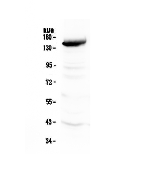
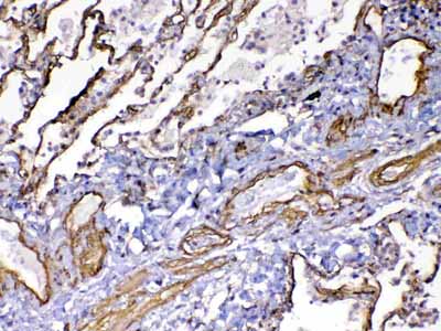
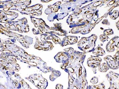
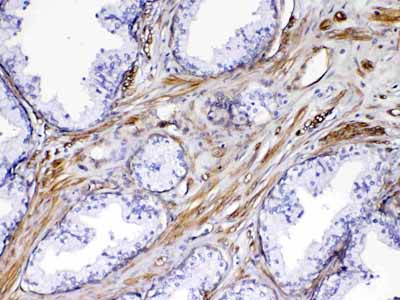
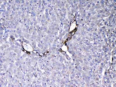
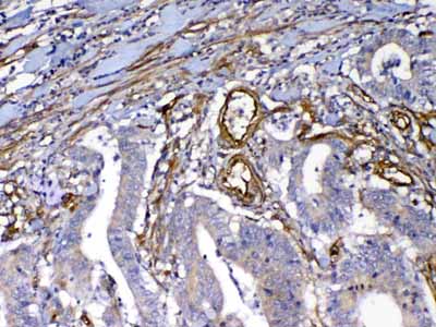
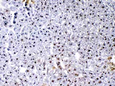
| Description | Rabbit IgG polyclonal antibody for VEGF Receptor 3 detection. Tested with WB, IHC-P, Direct ELISA in Human;Mouse;Rat. |
| Reconstitution | Add 0.2ml of distilled water will yield a concentration of 500ug/ml. |
| Gene ID | 2324 |
|---|---|
| Other Names | Vascular endothelial growth factor receptor 3, VEGFR-3, 2.7.10.1, Fms-like tyrosine kinase 4, FLT-4, Tyrosine-protein kinase receptor FLT4, FLT4, VEGFR3 |
| Calculated MW | 152757 Da |
| Application Details | Western blot, 0.1-0.5 µg/ml Immunohistochemistry(Paraffin-embedded Section), 0.5-1 µg/ml Direct ELISA, 0.1-0.5 µg/ml |
| Subcellular Localization | Cell membrane; Ligand-mediated autophosphorylation leads to rapid internalization. |
| Tissue Specificity | Detected in endothelial cells (at protein level). Widely expressed. Detected in fetal spleen, lung and brain. Detected in adult liver, muscle, thymus, placenta, lung, testis, ovary, prostate, heart, and kidney. |
| Contents | Each vial contains 4mg Trehalose, 0.9mg NaCl, 0.2mg Na2HPO4, 0.05mg NaN3. |
| Immunogen | E. coli-derived human VEGF Receptor 3 recombinant protein (Position: Y25-N259). |
| Cross Reactivity | No cross reactivity with other proteins. |
| Storage | At -20˚C; for one year. After r˚Constitution, at 4˚C; for one month. It˚Can also be aliquotted and stored frozen at -20˚C; for a longer time. Avoid repeated freezing and thawing. |
| Name | FLT4 |
|---|---|
| Synonyms | VEGFR3 |
| Function | Tyrosine-protein kinase that acts as a cell-surface receptor for VEGFC and VEGFD, and plays an essential role in adult lymphangiogenesis and in the development of the vascular network and the cardiovascular system during embryonic development. Promotes proliferation, survival and migration of endothelial cells, and regulates angiogenic sprouting. Signaling by activated FLT4 leads to enhanced production of VEGFC, and to a lesser degree VEGFA, thereby creating a positive feedback loop that enhances FLT4 signaling. Modulates KDR signaling by forming heterodimers. The secreted isoform 3 may function as a decoy receptor for VEGFC and/or VEGFD and play an important role as a negative regulator of VEGFC-mediated lymphangiogenesis and angiogenesis. Binding of vascular growth factors to isoform 1 or isoform 2 leads to the activation of several signaling cascades; isoform 2 seems to be less efficient in signal transduction, because it has a truncated C-terminus and therefore lacks several phosphorylation sites. Mediates activation of the MAPK1/ERK2, MAPK3/ERK1 signaling pathway, of MAPK8 and the JUN signaling pathway, and of the AKT1 signaling pathway. Phosphorylates SHC1. Mediates phosphorylation of PIK3R1, the regulatory subunit of phosphatidylinositol 3-kinase. Promotes phosphorylation of MAPK8 at 'Thr-183' and 'Tyr-185', and of AKT1 at 'Ser-473'. |
| Cellular Location | Cell membrane; Single-pass type I membrane protein Cytoplasm Nucleus. Note=Ligand-mediated autophosphorylation leads to rapid internalization [Isoform 2]: Cell membrane; Single-pass type I membrane protein |
| Tissue Location | Detected in endothelial cells (at protein level). Widely expressed. Detected in fetal spleen, lung and brain. Detected in adult liver, muscle, thymus, placenta, lung, testis, ovary, prostate, heart, and kidney. |

Thousands of laboratories across the world have published research that depended on the performance of antibodies from Abcepta to advance their research. Check out links to articles that cite our products in major peer-reviewed journals, organized by research category.
info@abcepta.com, and receive a free "I Love Antibodies" mug.

Provided below are standard protocols that you may find useful for product applications.
Background
Fms-related tyrosine kinase 4, also known as FLT4 or VEGFR3, is a protein which in humans is encoded by the FLT4 gene. It is mapped to 5q35.3. This gene encodes a tyrosine kinase receptor for vascular endothelial growth factors C and D. The protein is thought to be involved in lymphangiogenesis and maintenance of the lymphatic endothelium. FLT4 has an essential role in the development of the embryonic cardiovascular system before the emergence of the lymphatic vessels. It has been found that FLT4, which provides proangiogenic signaling when expressed on endothelium, may also have antiangiogenic properties when expressed at an avascular site by nonendothelial cells. FLT4 is also regarded as a regulator of vascular network formation.
If you have used an Abcepta product and would like to share how it has performed, please click on the "Submit Review" button and provide the requested information. Our staff will examine and post your review and contact you if needed.
If you have any additional inquiries please email technical services at tech@abcepta.com.
Ordering Information
Other Products
Shipping Information
