


 Foundational characteristics of cancer include proliferation, angiogenesis, migration, evasion of apoptosis, and cellular immortality. Find key markers for these cellular processes and antibodies to detect them.
 The SUMOplot™ Analysis Program predicts and scores sumoylation sites in your protein. SUMOylation is a post-translational modification involved in various cellular processes, such as nuclear-cytosolic transport, transcriptional regulation, apoptosis, protein stability, response to stress, and progression through the cell cycle.
 The Autophagy Receptor Motif Plotter predicts and scores autophagy receptor binding sites in your protein. Identifying proteins connected to this pathway is critical to understanding the role of autophagy in physiological as well as pathological processes such as development, differentiation, neurodegenerative diseases, stress, infection, and cancer.
Anti-VIP Receptor 1 Picoband Antibody
- SPECIFICATION
- CITATIONS
- PROTOCOLS
- BACKGROUND

Application
| WB |
|---|---|
| Primary Accession | P32241 |
| Host | Rabbit |
| Reactivity | Human, Mouse, Rat |
| Clonality | Polyclonal |
| Format | Lyophilized |
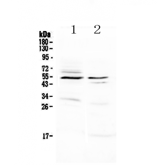
| Description | Rabbit IgG polyclonal antibody for VIP Receptor 1 detection. Tested with WB in Human;Mouse;Rat. |
| Reconstitution | Add 0.2ml of distilled water will yield a concentration of 500ug/ml. |
| Gene ID | 7433 |
|---|---|
| Other Names | Vasoactive intestinal polypeptide receptor 1, VIP-R-1, Pituitary adenylate cyclase-activating polypeptide type II receptor, PACAP type II receptor, PACAP-R-2, PACAP-R2, VPAC1, VIPR1 |
| Calculated MW | 51547 Da |
| Application Details | Western blot, 0.1-0.5 µg/ml |
| Subcellular Localization | Cell membrane; Multi-pass membrane protein. |
| Tissue Specificity | In lung, HT-29 colonic epithelial cells, Raji B-lymphoblasts. Lesser extent in brain, heart, kidney, liver and placenta. Not expressed in CD4+ or CD8+ T-cells. Expressed in the T-cell lines HARRIS, HuT 78, Jurkat and SUP-T1, but not in the T- cell lines Peer, MOLT-4, HSB and YT. |
| Contents | Each vial contains 4mg Trehalose, 0.9mg NaCl, 0.2mg Na2HPO4, 0.05mg NaN3. |
| Immunogen | A synthetic peptide corresponding to a sequence of human VIP Receptor 1 (QAELRRKWRRWHLQGVLGWNPKYRH). |
| Cross Reactivity | No cross reactivity with other proteins. |
| Storage | At -20˚C; for one year. After r˚Constitution, at 4˚C; for one month. It˚Can also be aliquotted and stored frozen at -20˚C; for a longer time. Avoid repeated freezing and thawing. |
| Name | VIPR1 (HGNC:12694) |
|---|---|
| Function | G protein-coupled receptor activated by the neuropeptides vasoactive intestinal peptide (VIP) and pituitary adenylate cyclase- activating polypeptide (ADCYAP1/PACAP) (PubMed:35477937, PubMed:36385145, PubMed:8179610). Binds VIP and both PACAP27 and PACAP38 bioactive peptides with the following order of ligand affinity VIP = PACAP27 > PACAP38 (PubMed:35477937, PubMed:8179610). Ligand binding causes a conformation change that triggers signaling via guanine nucleotide-binding proteins (G proteins) and modulates the activity of downstream effectors. Activates cAMP-dependent pathway (PubMed:35477937, PubMed:36385145, PubMed:8179610). |
| Cellular Location | Cell membrane; Multi-pass membrane protein |
| Tissue Location | In lung, HT-29 colonic epithelial cells, Raji B- lymphoblasts. Lesser extent in brain, heart, kidney, liver and placenta. Not expressed in CD4+ or CD8+ T-cells. Expressed in the T- cell lines HARRIS, HuT 78, Jurkat and SUP-T1, but not in the T-cell lines Peer, MOLT-4, HSB and YT. |

Thousands of laboratories across the world have published research that depended on the performance of antibodies from Abcepta to advance their research. Check out links to articles that cite our products in major peer-reviewed journals, organized by research category.
info@abcepta.com, and receive a free "I Love Antibodies" mug.

Provided below are standard protocols that you may find useful for product applications.
Background
VIPR1(Vasoactive intestinal polypeptide receptor 1),also known as VIPR,HVR1, is a protein that in humans is encoded by the VIPR1 gene. Distinct subsets of neural, respiratory, gastrointestinal, and immune cells bear specific high-affinity G protein-coupled receptors for VIP, such as VIPR1. The VIPR1 gene is mapped on 3p22.1. The VIPR1 gene was found to span approximately 22 kb and to be comprised of 13 exons (ranging from 42 to 1,400 bp) and 12 introns (ranging from 0.3 to 6.1 kb). One encodes a VIP receptor consisting of 460 amino acids and having 7 putative transmembrane domains, as do other G protein-coupled receptors. Patients with idiopathic achalasia show a significant difference in the distribution of SNPs affecting VIPR1.
If you have used an Abcepta product and would like to share how it has performed, please click on the "Submit Review" button and provide the requested information. Our staff will examine and post your review and contact you if needed.
If you have any additional inquiries please email technical services at tech@abcepta.com.
Ordering Information
Other Products
Shipping Information
