


 Foundational characteristics of cancer include proliferation, angiogenesis, migration, evasion of apoptosis, and cellular immortality. Find key markers for these cellular processes and antibodies to detect them.
 The SUMOplot™ Analysis Program predicts and scores sumoylation sites in your protein. SUMOylation is a post-translational modification involved in various cellular processes, such as nuclear-cytosolic transport, transcriptional regulation, apoptosis, protein stability, response to stress, and progression through the cell cycle.
 The Autophagy Receptor Motif Plotter predicts and scores autophagy receptor binding sites in your protein. Identifying proteins connected to this pathway is critical to understanding the role of autophagy in physiological as well as pathological processes such as development, differentiation, neurodegenerative diseases, stress, infection, and cancer.
> home > Products > Primary Antibodies > Signal Transduction > Anti-Phospho-YAP1 (S127) Rabbit Monoclonal Antibody
Anti-Phospho-YAP1 (S127) Rabbit Monoclonal Antibody
- SPECIFICATION
- CITATIONS
- PROTOCOLS
- BACKGROUND

Application
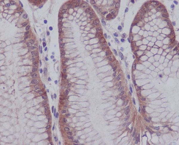
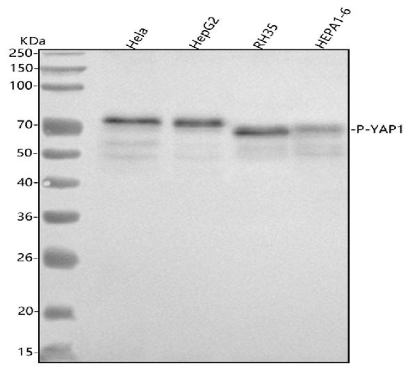
| WB, IHC |
|---|---|
| Primary Accession | P46937 |
| Host | Rabbit |
| Isotype | Rabbit IgG |
| Reactivity | Rat, Human, Mouse |
| Clonality | Monoclonal |
| Format | Liquid |
| Description | Anti-Phospho-YAP1 (S127) Rabbit Monoclonal Antibody . Tested in WB, IHC applications. This antibody reacts with Human, Mouse, Rat. |
| Gene ID | 10413 |
|---|---|
| Other Names | Transcriptional coactivator YAP1, Yes-associated protein 1, Protein yorkie homolog, Yes-associated protein YAP65 homolog, YAP1 (HGNC:16262), YAP65 |
| Calculated MW | 54462 MW KDa |
| Application Details | WB 1:5000-1:20000 IHC 1:50-1:200 |
| Subcellular Localization | Cytoplasm. Nucleus. Both phosphorylation and cell density can regulate its subcellular localization. Phosphorylation sequesters it in the cytoplasm by inhibiting its translocation into the nucleus. At low density, predominantly nuclear and is translocated to the cytoplasm at high density (PubMed:18158288, PubMed:20048001). PTPN14 induces translocation from the nucleus to the cytoplasm (PubMed:22525271).. |
| Tissue Specificity | Increased expression seen in some liver and prostate cancers. Isoforms lacking the transactivation domain found in striatal neurons of patients with Huntington disease (at protein level).. |
| Contents | Rabbit IgG in phosphate buffered saline, pH 7.4, 150mM NaCl, 0.02% sodium azide and 50% glycerol, 0.4-0.5mg/ml BSA. |
| Clone Names | Clone: GAB-25 |
| Immunogen | A synthesized peptide derived from human YAP1 |
| Purification | Affinity-chromatography |
| Storage | Store at -20°C for one year. For short term storage and frequent use, store at 4°C for up to one month. Avoid repeated freeze-thaw cycles. |
| Name | YAP1 (HGNC:16262) |
|---|---|
| Synonyms | YAP65 |
| Function | Transcriptional regulator with dual roles as a coactivator and corepressor. Critical downstream regulatory target in the Hippo signaling pathway, crucial for organ size control and tumor suppression by restricting proliferation and promoting apoptosis (PubMed:17974916, PubMed:18280240, PubMed:18579750, PubMed:21364637, PubMed:30447097). The Hippo signaling pathway core involves a kinase cascade featuring STK3/MST2 and STK4/MST1, along with its regulatory partner SAV1, which phosphorylates and activates LATS1/2 in complex with their regulatory protein, MOB1. This activation leads to the phosphorylation and inactivation of the YAP1 oncoprotein and WWTR1/TAZ (PubMed:18158288). Phosphorylation of YAP1 by LATS1/2 prevents its nuclear translocation, thereby regulating the expression of its target genes (PubMed:18158288, PubMed:26598551, PubMed:34404733). The transcriptional regulation of gene expression requires TEAD transcription factors and modulates cell growth, anchorage-independent growth, and induction of epithelial- mesenchymal transition (EMT) (PubMed:18579750). Plays a key role in tissue tension and 3D tissue shape by regulating the cortical actomyosin network, acting via ARHGAP18, a Rho GTPase activating protein that suppresses F-actin polymerization (PubMed:25778702). It also suppresses ciliogenesis by acting as a transcriptional corepressor of TEAD4 target genes AURKA and PLK1 (PubMed:25849865). In conjunction with WWTR1, regulates TGFB1-dependent SMAD2 and SMAD3 nuclear accumulation (By similarity). Synergizes with WBP2 to enhance PGR activity (PubMed:16772533). |
| Cellular Location | Cytoplasm. Nucleus. Cell junction, tight junction {ECO:0000250|UniProtKB:A0A8C0NGY6}. Cell membrane. Note=Both phosphorylation and cell density can regulate its subcellular localization (PubMed:18158288, PubMed:20048001). Phosphorylation sequesters it in the cytoplasm by inhibiting its translocation into the nucleus (PubMed:18158288, PubMed:20048001, PubMed:34404733). At low density, predominantly nuclear and is translocated to the cytoplasm at high density (PubMed:18158288, PubMed:20048001, PubMed:25849865). PTPN14 induces translocation from the nucleus to the cytoplasm (PubMed:22525271). In the nucleus, phosphorylation by PRP4K induces nuclear exclusion (PubMed:29695716). Localized mainly to the nucleus in the early stages of embryo development with expression becoming evident in the cytoplasm at the blastocyst and epiblast stages (By similarity) Localizes to the cytoplasm and tight junctions following interaction with AMOT isoform 1 (PubMed:21205866). Localizes to tight junctions following interaction with AMOTL2 (By similarity). Translocates to the nucleus in the presence of SNAIL1 (By similarity). Found at the cell membrane in keratinocytes in response to mechanical strain (PubMed:31835537). {ECO:0000250|UniProtKB:A0A8C0NGY6, ECO:0000250|UniProtKB:P46938, ECO:0000269|PubMed:18158288, ECO:0000269|PubMed:20048001, ECO:0000269|PubMed:21205866, ECO:0000269|PubMed:22525271, ECO:0000269|PubMed:25849865, ECO:0000269|PubMed:29695716, ECO:0000269|PubMed:31835537, ECO:0000269|PubMed:34404733} |
| Tissue Location | Increased expression seen in some liver and prostate cancers. Isoforms lacking the transactivation domain found in striatal neurons of patients with Huntington disease (at protein level). |
Research Areas
Citations (0)

Thousands of laboratories across the world have published research that depended on the performance of antibodies from Abcepta to advance their research. Check out links to articles that cite our products in major peer-reviewed journals, organized by research category.
Submit your citation using an Abcepta antibody to
info@abcepta.com, and receive a free "I Love Antibodies" mug.
info@abcepta.com, and receive a free "I Love Antibodies" mug.

Application Protocols
Provided below are standard protocols that you may find useful for product applications.
Abcepta welcomes feedback from its customers.
If you have used an Abcepta product and would like to share how it has performed, please click on the "Submit Review" button and provide the requested information. Our staff will examine and post your review and contact you if needed.
If you have any additional inquiries please email technical services at tech@abcepta.com.
$ 370.00
Cat# ABO13136
Ordering Information
United States
AlbaniaAustraliaAustriaBelgiumBosnia & HerzegovinaBrazilBulgariaCanadaCentral AmericaChinaCroatiaCyprusCzech RepublicDenmarkEstoniaFinlandFranceGermanyGreeceHong KongHungaryIcelandIndiaIndonesiaIrelandIsraelItalyJapanLatviaLithuaniaLuxembourgMacedoniaMalaysiaMaltaMexicoNetherlandsNew ZealandNorwayPakistanPolandPortugalRomaniaSerbiaSingaporeSlovakiaSloveniaSouth AfricaSouth KoreaSpainSwedenSwitzerlandTaiwanTurkeyUnited KingdomUnited StatesVietnamWorldwideOthers
USA Headquarters
(888) 735-7227 / (858) 622-0099 or (858) 875-1900
Other Products
Shipping Information
Domestic orders (in stock items)
Shipped out the same day. Orders placed after 1 PM (PST) will ship out the next business day.
International orders
Contact your local distributors
