


 Foundational characteristics of cancer include proliferation, angiogenesis, migration, evasion of apoptosis, and cellular immortality. Find key markers for these cellular processes and antibodies to detect them.
 The SUMOplot™ Analysis Program predicts and scores sumoylation sites in your protein. SUMOylation is a post-translational modification involved in various cellular processes, such as nuclear-cytosolic transport, transcriptional regulation, apoptosis, protein stability, response to stress, and progression through the cell cycle.
 The Autophagy Receptor Motif Plotter predicts and scores autophagy receptor binding sites in your protein. Identifying proteins connected to this pathway is critical to understanding the role of autophagy in physiological as well as pathological processes such as development, differentiation, neurodegenerative diseases, stress, infection, and cancer.
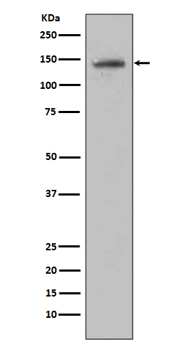
Anti-CD22/Siglec 2 Rabbit Monoclonal Antibody
- SPECIFICATION
- CITATIONS
- PROTOCOLS
- BACKGROUND

Application
| WB |
|---|---|
| Primary Accession | P20273 |
| Host | Rabbit |
| Isotype | Rabbit IgG |
| Reactivity | Rat, Human, Mouse |
| Clonality | Monoclonal |
| Format | Liquid |
| Description | Anti-CD22/Siglec 2 Rabbit Monoclonal Antibody . Tested in WB application. This antibody reacts with Human, Mouse, Rat. |
| Gene ID | 933 |
|---|---|
| Other Names | B-cell receptor CD22, B-lymphocyte cell adhesion molecule, BL-CAM, Sialic acid-binding Ig-like lectin 2, Siglec-2, T-cell surface antigen Leu-14, CD22, CD22 {ECO:0000303|PubMed:1691828, ECO:0000312|HGNC:HGNC:1643} |
| Calculated MW | 95348 MW KDa |
| Application Details | WB 1:500-1:2000 |
| Subcellular Localization | Cell membrane; Single-pass type I membrane protein. |
| Tissue Specificity | B-lymphocytes. |
| Contents | Rabbit IgG in phosphate buffered saline, pH 7.4, 150mM NaCl, 0.02% sodium azide and 50% glycerol, 0.4-0.5mg/ml BSA. |
| Clone Names | Clone: CBO-3 |
| Immunogen | A synthesized peptide derived from human CD22 |
| Purification | Affinity-chromatography |
| Storage | Store at -20°C for one year. For short term storage and frequent use, store at 4°C for up to one month. Avoid repeated freeze-thaw cycles. |
| Name | CD22 {ECO:0000303|PubMed:1691828, ECO:0000312|HGNC:HGNC:1643} |
|---|---|
| Function | Most highly expressed siglec (sialic acid-binding immunoglobulin-like lectin) on B-cells that plays a role in various aspects of B-cell biology including differentiation, antigen presentation, and trafficking to bone marrow (PubMed:34330755, PubMed:8627166). Binds to alpha 2,6-linked sialic acid residues of surface molecules such as CD22 itself, CD45 and IgM in a cis configuration. Can also bind to ligands on other cells as an adhesion molecule in a trans configuration (PubMed:20172905). Acts as an inhibitory coreceptor on the surface of B-cells and inhibits B-cell receptor induced signaling, characterized by inhibition of the calcium mobilization and cellular activation. Mechanistically, the immunoreceptor tyrosine-based inhibitory motif domain is phosphorylated by the Src kinase LYN, which in turn leads to the recruitment of the protein tyrosine phosphatase 1/PTPN6, leading to the negative regulation of BCR signaling (PubMed:8627166). If this negative signaling from is of sufficient strength, apoptosis of the B-cell can be induced (PubMed:20516366). |
| Cellular Location | Cell membrane; Single-pass type I membrane protein |
| Tissue Location | B-lymphocytes. |
Research Areas
Citations (0)

Thousands of laboratories across the world have published research that depended on the performance of antibodies from Abcepta to advance their research. Check out links to articles that cite our products in major peer-reviewed journals, organized by research category.
Submit your citation using an Abcepta antibody to
info@abcepta.com, and receive a free "I Love Antibodies" mug.
info@abcepta.com, and receive a free "I Love Antibodies" mug.

Application Protocols
Provided below are standard protocols that you may find useful for product applications.
Abcepta welcomes feedback from its customers.
If you have used an Abcepta product and would like to share how it has performed, please click on the "Submit Review" button and provide the requested information. Our staff will examine and post your review and contact you if needed.
If you have any additional inquiries please email technical services at tech@abcepta.com.
$ 370.00
Cat# ABO13347
Ordering Information
United States
AlbaniaAustraliaAustriaBelgiumBosnia & HerzegovinaBrazilBulgariaCanadaCentral AmericaChinaCroatiaCyprusCzech RepublicDenmarkEstoniaFinlandFranceGermanyGreeceHong KongHungaryIcelandIndiaIndonesiaIrelandIsraelItalyJapanLatviaLithuaniaLuxembourgMacedoniaMalaysiaMaltaMexicoNetherlandsNew ZealandNorwayPakistanPolandPortugalRomaniaSerbiaSingaporeSlovakiaSloveniaSouth AfricaSouth KoreaSpainSwedenSwitzerlandTaiwanTurkeyUnited KingdomUnited StatesVietnamWorldwideOthers
USA Headquarters
(888) 735-7227 / (858) 622-0099 or (858) 875-1900
Other Products
Shipping Information
Domestic orders (in stock items)
Shipped out the same day. Orders placed after 1 PM (PST) will ship out the next business day.
International orders
Contact your local distributors
