


 Foundational characteristics of cancer include proliferation, angiogenesis, migration, evasion of apoptosis, and cellular immortality. Find key markers for these cellular processes and antibodies to detect them.
 The SUMOplot™ Analysis Program predicts and scores sumoylation sites in your protein. SUMOylation is a post-translational modification involved in various cellular processes, such as nuclear-cytosolic transport, transcriptional regulation, apoptosis, protein stability, response to stress, and progression through the cell cycle.
 The Autophagy Receptor Motif Plotter predicts and scores autophagy receptor binding sites in your protein. Identifying proteins connected to this pathway is critical to understanding the role of autophagy in physiological as well as pathological processes such as development, differentiation, neurodegenerative diseases, stress, infection, and cancer.
> home > Products > Primary Antibodies > Antibody Collections > Head neck negative > Anti-CD59 Rabbit Monoclonal Antibody
Anti-CD59 Rabbit Monoclonal Antibody
- SPECIFICATION
- CITATIONS
- PROTOCOLS
- BACKGROUND

Application
| WB, IF, ICC, IP, FC |
|---|---|
| Primary Accession | P13987 |
| Host | Rabbit |
| Isotype | Rabbit IgG |
| Reactivity | Human |
| Clonality | Monoclonal |
| Format | Liquid |
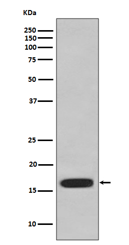
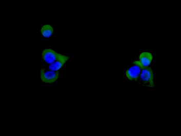
| Description | Anti-CD59 Rabbit Monoclonal Antibody . Tested in WB, ICC/IF, IP, Flow Cytometry applications. This antibody reacts with Human. |
| Gene ID | 966 |
|---|---|
| Other Names | CD59 glycoprotein, 1F5 antigen, 20 kDa homologous restriction factor, HRF-20, HRF20, MAC-inhibitory protein, MAC-IP, MEM43 antigen, Membrane attack complex inhibition factor, MACIF, Membrane inhibitor of reactive lysis, MIRL, Protectin, CD59, CD59, MIC11, MIN1, MIN2, MIN3, MSK21 |
| Calculated MW | 14177 MW KDa |
| Application Details | WB 1:1000-1:5000 ICC/IF 1:50-1:200 IP 1:50 FC 1:50 |
| Subcellular Localization | Cell membrane; Lipid-anchor, GPI-anchor. Secreted. Soluble form found in a number of tissues. |
| Contents | Rabbit IgG in phosphate buffered saline, pH 7.4, 150mM NaCl, 0.02% sodium azide and 50% glycerol, 0.4-0.5mg/ml BSA. |
| Clone Names | Clone: AABE-3 |
| Immunogen | A synthesized peptide derived from human CD59 |
| Purification | Affinity-chromatography |
| Storage | Store at -20°C for one year. For short term storage and frequent use, store at 4°C for up to one month. Avoid repeated freeze-thaw cycles. |
| Name | CD59 {ECO:0000303|PubMed:2475570, ECO:0000312|HGNC:HGNC:1689} |
|---|---|
| Function | Potent inhibitor of the complement membrane attack complex (MAC) action, which protects human cells from damage during complement activation (PubMed:11882685, PubMed:1698710, PubMed:2475111, PubMed:2475570, PubMed:2606909, PubMed:9053451). Acts by binding to the beta-haipins of C8 (C8A and C8B) components of the assembling MAC, forming an intermolecular beta-sheet that prevents incorporation of the multiple copies of C9 required for complete formation of the osmolytic pore (PubMed:11882685, PubMed:1698710, PubMed:36797260). |
| Cellular Location | Cell membrane; Lipid-anchor, GPI-anchor. Secreted. Note=Localizes to the cell surface (PubMed:36797260). Soluble form found in a number of tissues (PubMed:8670172). |
Research Areas
Citations (0)

Thousands of laboratories across the world have published research that depended on the performance of antibodies from Abcepta to advance their research. Check out links to articles that cite our products in major peer-reviewed journals, organized by research category.
Submit your citation using an Abcepta antibody to
info@abcepta.com, and receive a free "I Love Antibodies" mug.
info@abcepta.com, and receive a free "I Love Antibodies" mug.

Application Protocols
Provided below are standard protocols that you may find useful for product applications.
Abcepta welcomes feedback from its customers.
If you have used an Abcepta product and would like to share how it has performed, please click on the "Submit Review" button and provide the requested information. Our staff will examine and post your review and contact you if needed.
If you have any additional inquiries please email technical services at tech@abcepta.com.
$ 185.00

⚠️ Limit: 5 vials per order for discount.
Cat# ABO13527
Ordering Information
United States
AlbaniaAustraliaAustriaBelgiumBosnia & HerzegovinaBrazilBulgariaCanadaCentral AmericaChinaCroatiaCyprusCzech RepublicDenmarkEstoniaFinlandFranceGermanyGreeceHong KongHungaryIcelandIndiaIndonesiaIrelandIsraelItalyJapanLatviaLithuaniaLuxembourgMacedoniaMalaysiaMaltaMexicoNetherlandsNew ZealandNorwayPakistanPolandPortugalRomaniaSerbiaSingaporeSlovakiaSloveniaSouth AfricaSouth KoreaSpainSwedenSwitzerlandTaiwanTurkeyUnited KingdomUnited StatesVietnamWorldwideOthers
USA Headquarters
(888) 735-7227 / (858) 622-0099 or (858) 875-1900
Other Products
Shipping Information
Domestic orders (in stock items)
Shipped out the same day. Orders placed after 1 PM (PST) will ship out the next business day.
International orders
Contact your local distributors
