


 Foundational characteristics of cancer include proliferation, angiogenesis, migration, evasion of apoptosis, and cellular immortality. Find key markers for these cellular processes and antibodies to detect them.
 The SUMOplot™ Analysis Program predicts and scores sumoylation sites in your protein. SUMOylation is a post-translational modification involved in various cellular processes, such as nuclear-cytosolic transport, transcriptional regulation, apoptosis, protein stability, response to stress, and progression through the cell cycle.
 The Autophagy Receptor Motif Plotter predicts and scores autophagy receptor binding sites in your protein. Identifying proteins connected to this pathway is critical to understanding the role of autophagy in physiological as well as pathological processes such as development, differentiation, neurodegenerative diseases, stress, infection, and cancer.
> home > Products > Primary Antibodies > Signal Transduction > Anti-NFAT1 NFATC2 Rabbit Monoclonal Antibody
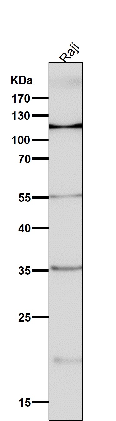
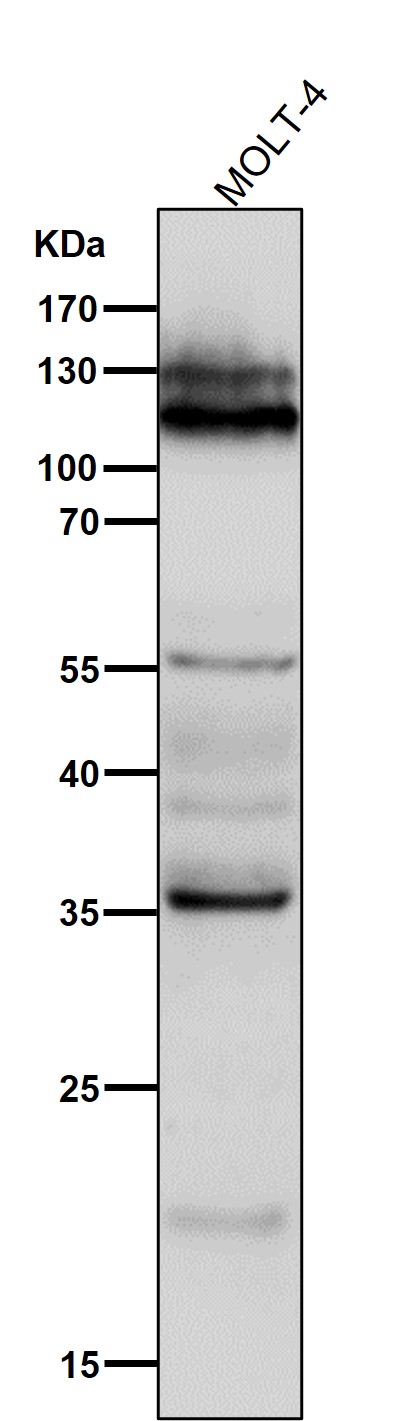
Anti-NFAT1 NFATC2 Rabbit Monoclonal Antibody

- SPECIFICATION
- CITATIONS
- PROTOCOLS
- BACKGROUND

Application
| WB |
|---|---|
| Primary Accession | Q13469 |
| Host | Rabbit |
| Isotype | Rabbit IgG |
| Reactivity | Human |
| Clonality | Monoclonal |
| Format | Liquid |
| Description | Anti-NFAT1 NFATC2 Rabbit Monoclonal Antibody . Tested in WB application. This antibody reacts with Human. |
| Gene ID | 4773 |
|---|---|
| Other Names | Nuclear factor of activated T-cells, cytoplasmic 2, NF-ATc2, NFATc2, NFAT pre-existing subunit, NF-ATp, T-cell transcription factor NFAT1, NFATC2, NFAT1, NFATP |
| Calculated MW | 100146 MW KDa |
| Application Details | WB 1:500-1:2000 |
| Subcellular Localization | Cytoplasm. Nucleus. Cytoplasmic for the phosphorylated form and nuclear after activation that is controlled by calcineurin-mediated dephosphorylation. Rapid nuclear exit of NFATC is thought to be one mechanism by which cells distinguish between sustained and transient calcium signals. The subcellular localization of NFATC plays a key role in the regulation of gene transcription. |
| Tissue Specificity | Expressed in thymus, spleen, heart, testis, brain, placenta, muscle and pancreas. Isoform 1 is highly expressed in the small intestine, heart, testis, prostate, thymus, placenta and thyroid. Isoform 3 is highly expressed in stomach, uterus, placenta, trachea and thyroid.. |
| Contents | Rabbit IgG in phosphate buffered saline, pH 7.4, 150mM NaCl, 0.02% sodium azide and 50% glycerol, 0.4-0.5mg/ml BSA. |
| Clone Names | Clone: AAFG-14 |
| Immunogen | A synthesized peptide derived from human NFAT1 |
| Purification | Affinity-chromatography |
| Storage | Store at -20°C for one year. For short term storage and frequent use, store at 4°C for up to one month. Avoid repeated freeze-thaw cycles. |
| Name | NFATC2 |
|---|---|
| Synonyms | NFAT1, NFATP |
| Function | Plays a role in the inducible expression of cytokine genes in T-cells, especially in the induction of the IL-2, IL-3, IL-4, TNF-alpha or GM-CSF (PubMed:15790681). Promotes invasive migration through the activation of GPC6 expression and WNT5A signaling pathway (PubMed:21871017). Is involved in the negative regulation of chondrogenesis (PubMed:35789258). Recruited by AKAP5 to ORAI1 pore- forming subunit of CRAC channels in Ca(2+) signaling microdomains where store-operated Ca(2+) influx is coupled to calmodulin and calcineurin signaling and activation of NFAT-dependent transcriptional responses. |
| Cellular Location | Cytoplasm. Nucleus. Note=Cytoplasmic for the phosphorylated form and nuclear after activation that is controlled by calcineurin-mediated dephosphorylation. Rapid nuclear exit of NFATC is thought to be one mechanism by which cells distinguish between sustained and transient calcium signals. The subcellular localization of NFATC plays a key role in the regulation of gene transcription |
| Tissue Location | Expressed in thymus, spleen, heart, testis, brain, placenta, muscle and pancreas. Isoform 1 is highly expressed in the small intestine, heart, testis, prostate, thymus, placenta and thyroid Isoform 3 is highly expressed in stomach, uterus, placenta, trachea and thyroid. |
Research Areas
Citations (0)

Thousands of laboratories across the world have published research that depended on the performance of antibodies from Abcepta to advance their research. Check out links to articles that cite our products in major peer-reviewed journals, organized by research category.
Submit your citation using an Abcepta antibody to
info@abcepta.com, and receive a free "I Love Antibodies" mug.
info@abcepta.com, and receive a free "I Love Antibodies" mug.

Application Protocols
Provided below are standard protocols that you may find useful for product applications.
Abcepta welcomes feedback from its customers.
If you have used an Abcepta product and would like to share how it has performed, please click on the "Submit Review" button and provide the requested information. Our staff will examine and post your review and contact you if needed.
If you have any additional inquiries please email technical services at tech@abcepta.com.
$ 370.00
Cat# ABO13772
Ordering Information
United States
AlbaniaAustraliaAustriaBelgiumBosnia & HerzegovinaBrazilBulgariaCanadaCentral AmericaChinaCroatiaCyprusCzech RepublicDenmarkEstoniaFinlandFranceGermanyGreeceHong KongHungaryIcelandIndiaIndonesiaIrelandIsraelItalyJapanLatviaLithuaniaLuxembourgMacedoniaMalaysiaMaltaMexicoNetherlandsNew ZealandNorwayPakistanPolandPortugalRomaniaSerbiaSingaporeSlovakiaSloveniaSouth AfricaSouth KoreaSpainSwedenSwitzerlandTaiwanTurkeyUnited KingdomUnited StatesVietnamWorldwideOthers
USA Headquarters
(888) 735-7227 / (858) 622-0099 or (858) 875-1900
Other Products
Shipping Information
Domestic orders (in stock items)
Shipped out the same day. Orders placed after 1 PM (PST) will ship out the next business day.
International orders
Contact your local distributors
