


 Foundational characteristics of cancer include proliferation, angiogenesis, migration, evasion of apoptosis, and cellular immortality. Find key markers for these cellular processes and antibodies to detect them.
 The SUMOplot™ Analysis Program predicts and scores sumoylation sites in your protein. SUMOylation is a post-translational modification involved in various cellular processes, such as nuclear-cytosolic transport, transcriptional regulation, apoptosis, protein stability, response to stress, and progression through the cell cycle.
 The Autophagy Receptor Motif Plotter predicts and scores autophagy receptor binding sites in your protein. Identifying proteins connected to this pathway is critical to understanding the role of autophagy in physiological as well as pathological processes such as development, differentiation, neurodegenerative diseases, stress, infection, and cancer.
> home > Products > Primary Antibodies > Antibody Collections > Ovary positive > Anti-Sumo 1 Rabbit Monoclonal Antibody
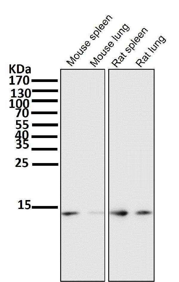
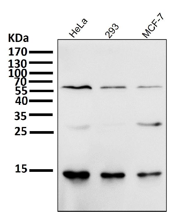
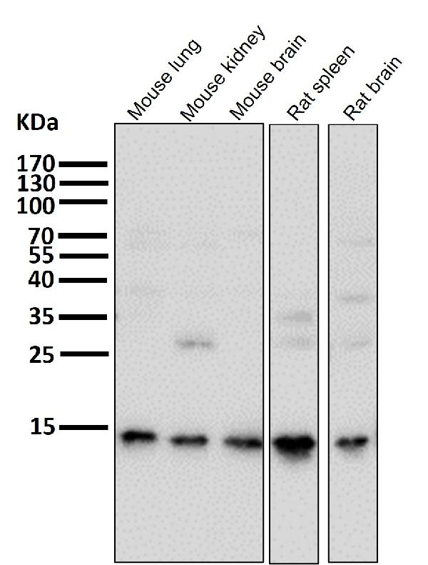
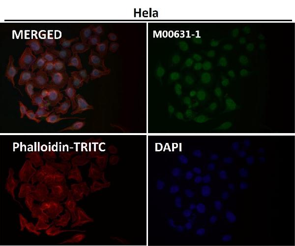
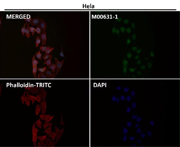
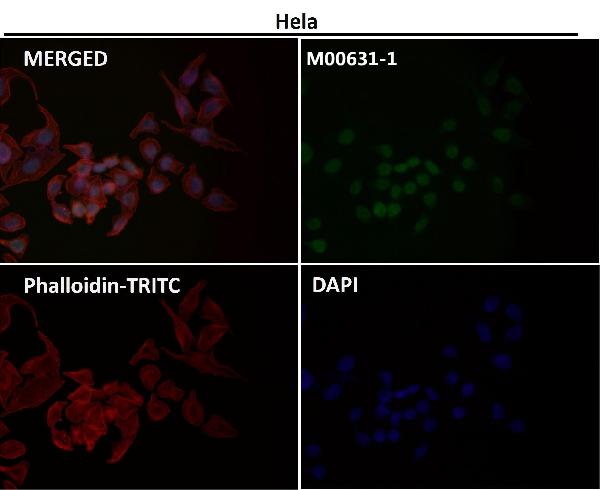
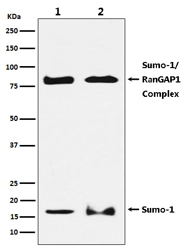
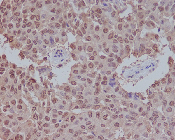
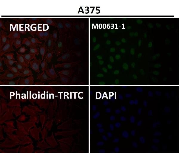
Anti-Sumo 1 Rabbit Monoclonal Antibody


- SPECIFICATION
- CITATIONS
- PROTOCOLS
- BACKGROUND

Application
| WB, IHC, IF, ICC, IP, FC |
|---|---|
| Primary Accession | P63165 |
| Host | Rabbit |
| Isotype | Rabbit IgG |
| Reactivity | Rat, Human, Mouse |
| Clonality | Monoclonal |
| Format | Liquid |
| Description | Anti-Sumo 1 Rabbit Monoclonal Antibody . Tested in WB, IHC, ICC/IF, IP, Flow Cytometry applications. This antibody reacts with Human, Mouse, Rat. |
| Gene ID | 7341 |
|---|---|
| Other Names | Small ubiquitin-related modifier 1, SUMO-1, GAP-modifying protein 1, GMP1, SMT3 homolog 3, Sentrin, Ubiquitin-homology domain protein PIC1, Ubiquitin-like protein SMT3C, Smt3C, Ubiquitin-like protein UBL1, SUMO1, SMT3C, SMT3H3, UBL1 |
| Calculated MW | 11557 MW KDa |
| Application Details | WB 1:500-1:2000 IHC 1:50-1:200 ICC/IF 1:50-1:200 IP 1:50 FC 1:50 |
| Subcellular Localization | Nucleus membrane. Nucleus speckle. Cytoplasm. Nucleus, PML body. Cell membrane. Recruited by BCL11A into the nuclear body.. |
| Contents | Rabbit IgG in phosphate buffered saline, pH 7.4, 150mM NaCl, 0.02% sodium azide and 50% glycerol, 0.4-0.5mg/ml BSA. |
| Clone Names | Clone: BFI-19 |
| Immunogen | A synthesized peptide derived from human Sumo 1 |
| Purification | Affinity-chromatography |
| Storage | Store at -20°C for one year. For short term storage and frequent use, store at 4°C for up to one month. Avoid repeated freeze-thaw cycles. |
| Name | SUMO1 |
|---|---|
| Synonyms | SMT3C, SMT3H3, UBL1 |
| Function | Ubiquitin-like protein that can be covalently attached to proteins as a monomer or a lysine-linked polymer. Covalent attachment via an isopeptide bond to its substrates requires prior activation by the E1 complex SAE1-SAE2 and linkage to the E2 enzyme UBE2I, and can be promoted by E3 ligases such as PIAS1-4, RANBP2 or CBX4. This post- translational modification on lysine residues of proteins plays a crucial role in a number of cellular processes such as nuclear transport, DNA replication and repair, mitosis and signal transduction. Involved for instance in targeting RANGAP1 to the nuclear pore complex protein RANBP2. Covalently attached to the voltage-gated potassium channel KCNB1; this modulates the gating characteristics of KCNB1 (PubMed:19223394). Polymeric SUMO1 chains are also susceptible to polyubiquitination which functions as a signal for proteasomal degradation of modified proteins. May also regulate a network of genes involved in palate development. Covalently attached to ZFHX3 (PubMed:24651376). |
| Cellular Location | Nucleus membrane. Nucleus speckle {ECO:0000250|UniProtKB:P63166}. Cytoplasm. Nucleus, PML body. Cell membrane. Nucleus. Note=Recruited by BCL11A into the nuclear body (By similarity). In the presence of ZFHX3, sequesterd to nuclear body (NB)-like dots in the nucleus some of which overlap or closely associate with PML body (PubMed:24651376) {ECO:0000250|UniProtKB:P63166, ECO:0000269|PubMed:24651376} |
Research Areas
Citations (0)

Thousands of laboratories across the world have published research that depended on the performance of antibodies from Abcepta to advance their research. Check out links to articles that cite our products in major peer-reviewed journals, organized by research category.
Submit your citation using an Abcepta antibody to
info@abcepta.com, and receive a free "I Love Antibodies" mug.
info@abcepta.com, and receive a free "I Love Antibodies" mug.

Application Protocols
Provided below are standard protocols that you may find useful for product applications.
Abcepta welcomes feedback from its customers.
If you have used an Abcepta product and would like to share how it has performed, please click on the "Submit Review" button and provide the requested information. Our staff will examine and post your review and contact you if needed.
If you have any additional inquiries please email technical services at tech@abcepta.com.
$ 370.00
Cat# ABO13822
Ordering Information
United States
AlbaniaAustraliaAustriaBelgiumBosnia & HerzegovinaBrazilBulgariaCanadaCentral AmericaChinaCroatiaCyprusCzech RepublicDenmarkEstoniaFinlandFranceGermanyGreeceHong KongHungaryIcelandIndiaIndonesiaIrelandIsraelItalyJapanLatviaLithuaniaLuxembourgMacedoniaMalaysiaMaltaMexicoNetherlandsNew ZealandNorwayPakistanPolandPortugalRomaniaSerbiaSingaporeSlovakiaSloveniaSouth AfricaSouth KoreaSpainSwedenSwitzerlandTaiwanTurkeyUnited KingdomUnited StatesVietnamWorldwideOthers
USA Headquarters
(888) 735-7227 / (858) 622-0099 or (858) 875-1900
Other Products
Shipping Information
Domestic orders (in stock items)
Shipped out the same day. Orders placed after 1 PM (PST) will ship out the next business day.
International orders
Contact your local distributors
