


 Foundational characteristics of cancer include proliferation, angiogenesis, migration, evasion of apoptosis, and cellular immortality. Find key markers for these cellular processes and antibodies to detect them.
 The SUMOplot™ Analysis Program predicts and scores sumoylation sites in your protein. SUMOylation is a post-translational modification involved in various cellular processes, such as nuclear-cytosolic transport, transcriptional regulation, apoptosis, protein stability, response to stress, and progression through the cell cycle.
 The Autophagy Receptor Motif Plotter predicts and scores autophagy receptor binding sites in your protein. Identifying proteins connected to this pathway is critical to understanding the role of autophagy in physiological as well as pathological processes such as development, differentiation, neurodegenerative diseases, stress, infection, and cancer.
Anti-MINA53 Rabbit Monoclonal Antibody
- SPECIFICATION
- CITATIONS
- PROTOCOLS
- BACKGROUND

Application
| WB, IF, ICC |
|---|---|
| Primary Accession | Q8IUF8 |
| Host | Rabbit |
| Isotype | Rabbit IgG |
| Reactivity | Human, Mouse |
| Clonality | Monoclonal |
| Format | Liquid |
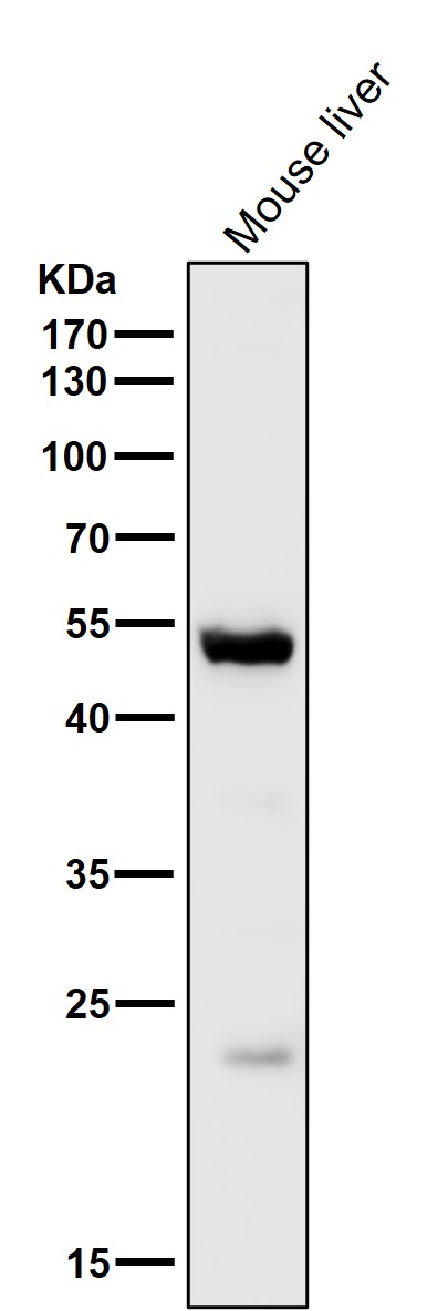
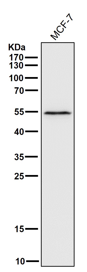
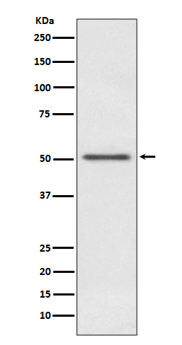
| Description | Anti-MINA53 Rabbit Monoclonal Antibody . Tested in WB, ICC/IF applications. This antibody reacts with Human, Mouse. |
| Gene ID | 84864 |
|---|---|
| Other Names | Ribosomal oxygenase 2 {ECO:0000312|HGNC:HGNC:19441}, 60S ribosomal protein L27a histidine hydroxylase, Bifunctional lysine-specific demethylase and histidyl-hydroxylase MINA, 1.14.11.79, Histone lysine demethylase MINA, MYC-induced nuclear antigen, Mineral dust-induced gene protein, Nucleolar protein 52, Ribosomal oxygenase MINA, ROX, RIOX2 (HGNC:19441) |
| Calculated MW | 52800 MW KDa |
| Application Details | WB 1:500-1:2000 ICC/IF 1:50-1:200 |
| Subcellular Localization | Nucleus. Nucleus, nucleolus. |
| Tissue Specificity | Expressed in liver, skeletal muscle, heart, pancreas, and placenta. Not detected in brain, lung or kidney. Expressed in several lung cancer tissues, but is barely detected in the adjacent non-cancerous tissues. Also highly expressed in several esophageal squamous cell carcinoma (ESCC), and colon cancer tissues, and in various cancer cell lines.. |
| Contents | Rabbit IgG in phosphate buffered saline, pH 7.4, 150mM NaCl, 0.02% sodium azide and 50% glycerol, 0.4-0.5mg/ml BSA. |
| Clone Names | Clone: AAAO-13 |
| Immunogen | A synthesized peptide derived from human MINA53 |
| Purification | Affinity-chromatography |
| Storage | Store at -20°C for one year. For short term storage and frequent use, store at 4°C for up to one month. Avoid repeated freeze-thaw cycles. |
| Name | RIOX2 (HGNC:19441) |
|---|---|
| Function | Oxygenase that can act as both a histone lysine demethylase and a ribosomal histidine hydroxylase. Is involved in the demethylation of trimethylated 'Lys-9' on histone H3 (H3K9me3), leading to an increase in ribosomal RNA expression. Also catalyzes the hydroxylation of 60S ribosomal protein L27a on 'His-39'. May play an important role in cell growth and survival. May be involved in ribosome biogenesis, most likely during the assembly process of pre-ribosomal particles. |
| Cellular Location | Nucleus. Nucleus, nucleolus |
| Tissue Location | Expressed in liver, skeletal muscle, heart, pancreas, and placenta. Not detected in brain, lung or kidney Expressed in several lung cancer tissues, but is barely detected in the adjacent non-cancerous tissues. Also highly expressed in several esophageal squamous cell carcinoma (ESCC), and colon cancer tissues, and in various cancer cell lines. |
Citations (0)

Thousands of laboratories across the world have published research that depended on the performance of antibodies from Abcepta to advance their research. Check out links to articles that cite our products in major peer-reviewed journals, organized by research category.
Submit your citation using an Abcepta antibody to
info@abcepta.com, and receive a free "I Love Antibodies" mug.
info@abcepta.com, and receive a free "I Love Antibodies" mug.

Application Protocols
Provided below are standard protocols that you may find useful for product applications.
Abcepta welcomes feedback from its customers.
If you have used an Abcepta product and would like to share how it has performed, please click on the "Submit Review" button and provide the requested information. Our staff will examine and post your review and contact you if needed.
If you have any additional inquiries please email technical services at tech@abcepta.com.
$ 370.00
Cat# ABO13867
Ordering Information
United States
AlbaniaAustraliaAustriaBelgiumBosnia & HerzegovinaBrazilBulgariaCanadaCentral AmericaChinaCroatiaCyprusCzech RepublicDenmarkEstoniaFinlandFranceGermanyGreeceHong KongHungaryIcelandIndiaIndonesiaIrelandIsraelItalyJapanLatviaLithuaniaLuxembourgMacedoniaMalaysiaMaltaMexicoNetherlandsNew ZealandNorwayPakistanPolandPortugalRomaniaSerbiaSingaporeSlovakiaSloveniaSouth AfricaSouth KoreaSpainSwedenSwitzerlandTaiwanTurkeyUnited KingdomUnited StatesVietnamWorldwideOthers
USA Headquarters
(888) 735-7227 / (858) 622-0099 or (858) 875-1900
Other Products
Shipping Information
Domestic orders (in stock items)
Shipped out the same day. Orders placed after 1 PM (PST) will ship out the next business day.
International orders
Contact your local distributors
