


 Foundational characteristics of cancer include proliferation, angiogenesis, migration, evasion of apoptosis, and cellular immortality. Find key markers for these cellular processes and antibodies to detect them.
 The SUMOplot™ Analysis Program predicts and scores sumoylation sites in your protein. SUMOylation is a post-translational modification involved in various cellular processes, such as nuclear-cytosolic transport, transcriptional regulation, apoptosis, protein stability, response to stress, and progression through the cell cycle.
 The Autophagy Receptor Motif Plotter predicts and scores autophagy receptor binding sites in your protein. Identifying proteins connected to this pathway is critical to understanding the role of autophagy in physiological as well as pathological processes such as development, differentiation, neurodegenerative diseases, stress, infection, and cancer.
> home > Products > Primary Antibodies > Signal Transduction > Anti-ATP5A1/Atp5A Rabbit Monoclonal Antibody
Anti-ATP5A1/Atp5A Rabbit Monoclonal Antibody
- SPECIFICATION
- CITATIONS
- PROTOCOLS
- BACKGROUND

Application
| WB, IHC, IF, ICC, FC |
|---|---|
| Primary Accession | P25705 |
| Host | Rabbit |
| Isotype | Rabbit IgG |
| Reactivity | Rat, Human, Mouse |
| Clonality | Monoclonal |
| Format | Liquid |
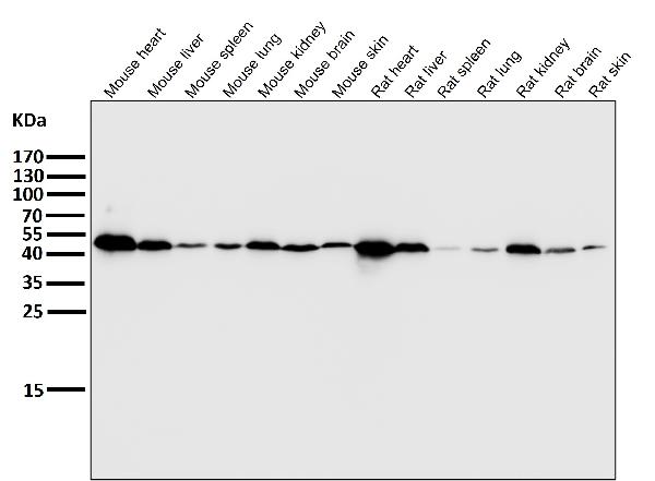
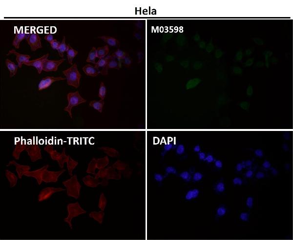
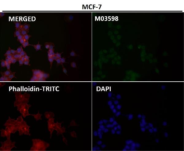
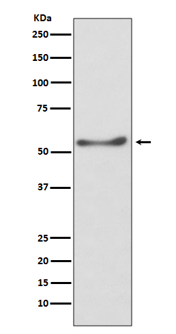
| Description | Anti-ATP5A1/Atp5A Rabbit Monoclonal Antibody . Tested in WB, IHC, ICC/IF, Flow Cytometry applications. This antibody reacts with Human, Mouse, Rat. |
| Gene ID | 498 |
|---|---|
| Other Names | ATP synthase subunit alpha, mitochondrial, ATP synthase F1 subunit alpha {ECO:0000312|HGNC:HGNC:823}, ATP5F1A (HGNC:823) |
| Calculated MW | 59751 MW KDa |
| Application Details | WB 1:500-1:2000 IHC 1:50-1:200 ICC/IF 1:50-1:200 FC 1:50 |
| Subcellular Localization | Mitochondrion inner membrane. Cell membrane; Peripheral membrane protein; Extracellular side. Colocalizes with HRG on the cell surface of T-cells (PubMed:19285951). |
| Tissue Specificity | Fetal lung, heart, liver, gut and kidney. Expressed at higher levels in the fetal brain, retina and spinal cord.. |
| Contents | Rabbit IgG in phosphate buffered saline, pH 7.4, 150mM NaCl, 0.02% sodium azide and 50% glycerol, 0.4-0.5mg/ml BSA. |
| Clone Names | Clone: AAFC-1 |
| Immunogen | A synthesized peptide derived from human ATP5A1 |
| Purification | Affinity-chromatography |
| Storage | Store at -20°C for one year. For short term storage and frequent use, store at 4°C for up to one month. Avoid repeated freeze-thaw cycles. |
| Name | ATP5F1A (HGNC:823) |
|---|---|
| Function | Subunit alpha, of the mitochondrial membrane ATP synthase complex (F(1)F(0) ATP synthase or Complex V) that produces ATP from ADP in the presence of a proton gradient across the membrane which is generated by electron transport complexes of the respiratory chain (Probable). ATP synthase complex consist of a soluble F(1) head domain - the catalytic core - and a membrane F(1) domain - the membrane proton channel (PubMed:37244256). These two domains are linked by a central stalk rotating inside the F(1) region and a stationary peripheral stalk (PubMed:37244256). During catalysis, ATP synthesis in the catalytic domain of F(1) is coupled via a rotary mechanism of the central stalk subunits to proton translocation (Probable). In vivo, can only synthesize ATP although its ATP hydrolase activity can be activated artificially in vitro (By similarity). With the catalytic subunit beta (ATP5F1B), forms the catalytic core in the F(1) domain (PubMed:37244256). Subunit alpha does not bear the catalytic high- affinity ATP-binding sites (Probable). Binds the bacterial siderophore enterobactin and can promote mitochondrial accumulation of enterobactin-derived iron ions (PubMed:30146159). |
| Cellular Location | Mitochondrion. Mitochondrion inner membrane {ECO:0000250|UniProtKB:P19483}; Peripheral membrane protein {ECO:0000250|UniProtKB:P19483}; Matrix side {ECO:0000250|UniProtKB:P19483}. Cell membrane; Peripheral membrane protein; Extracellular side. Note=Colocalizes with HRG on the cell surface of T-cells (PubMed:19285951). |
| Tissue Location | Fetal lung, heart, liver, gut and kidney. Expressed at higher levels in the fetal brain, retina and spinal cord |
Research Areas
Citations (0)

Thousands of laboratories across the world have published research that depended on the performance of antibodies from Abcepta to advance their research. Check out links to articles that cite our products in major peer-reviewed journals, organized by research category.
Submit your citation using an Abcepta antibody to
info@abcepta.com, and receive a free "I Love Antibodies" mug.
info@abcepta.com, and receive a free "I Love Antibodies" mug.

Application Protocols
Provided below are standard protocols that you may find useful for product applications.
Abcepta welcomes feedback from its customers.
If you have used an Abcepta product and would like to share how it has performed, please click on the "Submit Review" button and provide the requested information. Our staff will examine and post your review and contact you if needed.
If you have any additional inquiries please email technical services at tech@abcepta.com.
$ 370.00
Cat# ABO13871
Ordering Information
United States
AlbaniaAustraliaAustriaBelgiumBosnia & HerzegovinaBrazilBulgariaCanadaCentral AmericaChinaCroatiaCyprusCzech RepublicDenmarkEstoniaFinlandFranceGermanyGreeceHong KongHungaryIcelandIndiaIndonesiaIrelandIsraelItalyJapanLatviaLithuaniaLuxembourgMacedoniaMalaysiaMaltaMexicoNetherlandsNew ZealandNorwayPakistanPolandPortugalRomaniaSerbiaSingaporeSlovakiaSloveniaSouth AfricaSouth KoreaSpainSwedenSwitzerlandTaiwanTurkeyUnited KingdomUnited StatesVietnamWorldwideOthers
USA Headquarters
(888) 735-7227 / (858) 622-0099 or (858) 875-1900
Other Products
Shipping Information
Domestic orders (in stock items)
Shipped out the same day. Orders placed after 1 PM (PST) will ship out the next business day.
International orders
Contact your local distributors
