


 Foundational characteristics of cancer include proliferation, angiogenesis, migration, evasion of apoptosis, and cellular immortality. Find key markers for these cellular processes and antibodies to detect them.
 The SUMOplot™ Analysis Program predicts and scores sumoylation sites in your protein. SUMOylation is a post-translational modification involved in various cellular processes, such as nuclear-cytosolic transport, transcriptional regulation, apoptosis, protein stability, response to stress, and progression through the cell cycle.
 The Autophagy Receptor Motif Plotter predicts and scores autophagy receptor binding sites in your protein. Identifying proteins connected to this pathway is critical to understanding the role of autophagy in physiological as well as pathological processes such as development, differentiation, neurodegenerative diseases, stress, infection, and cancer.
> home > Products > Primary Antibodies > Signal Transduction > Anti-CaMKII delta CAMK2D Rabbit Monoclonal Antibody
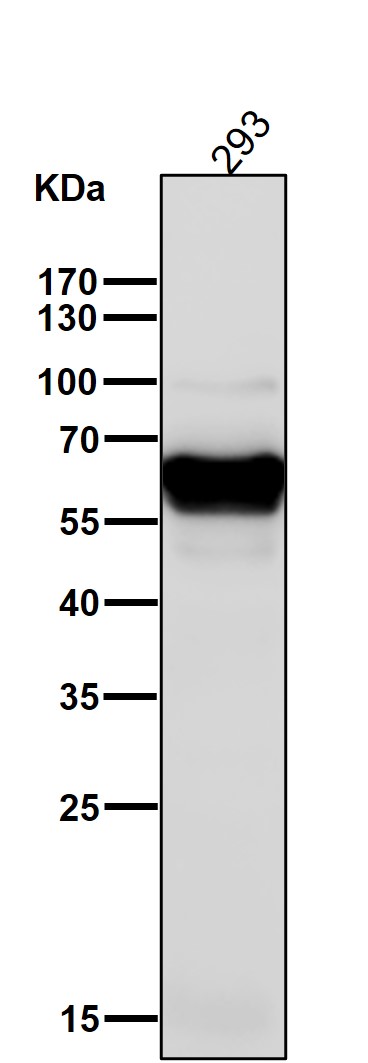
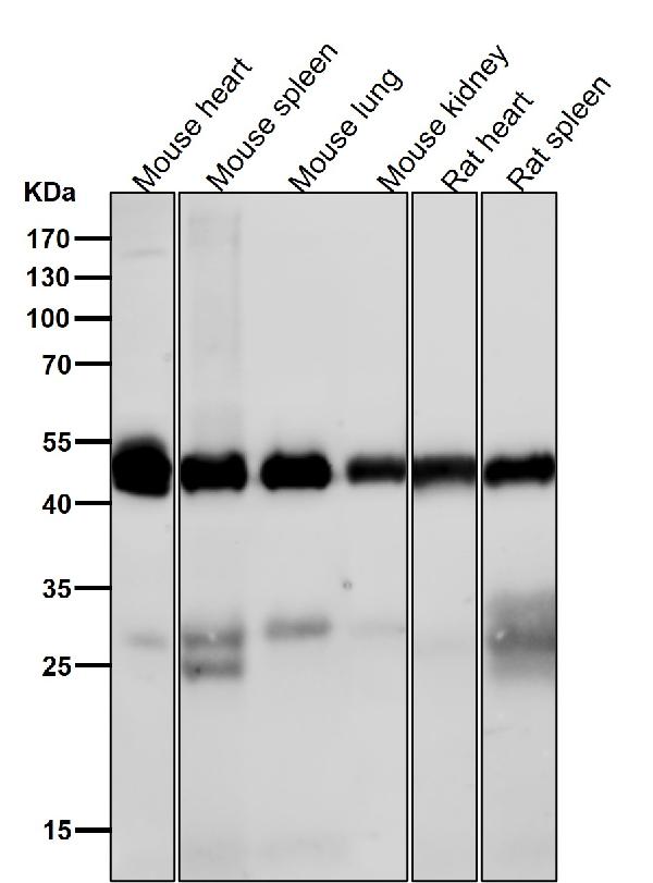
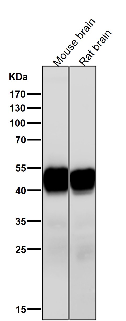
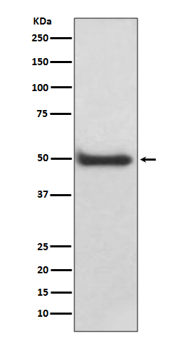
Anti-CaMKII delta CAMK2D Rabbit Monoclonal Antibody
- SPECIFICATION
- CITATIONS
- PROTOCOLS
- BACKGROUND

Application
| WB, IHC |
|---|---|
| Primary Accession | Q13557 |
| Host | Rabbit |
| Isotype | Rabbit IgG |
| Reactivity | Rat, Human, Mouse |
| Clonality | Monoclonal |
| Format | Liquid |
| Description | Anti-CaMKII delta CAMK2D Rabbit Monoclonal Antibody . Tested in WB, IHC applications. This antibody reacts with Human, Mouse, Rat. |
| Gene ID | 817 |
|---|---|
| Other Names | Calcium/calmodulin-dependent protein kinase type II subunit delta, CaM kinase II subunit delta, CaMK-II subunit delta, 2.7.11.17, CAMK2D, CAMKD |
| Calculated MW | 56369 MW KDa |
| Application Details | WB 1:500-1:2000 IHC 1:50-1:200 |
| Subcellular Localization | Cell membrane, sarcolemma ; Peripheral membrane protein ; Cytoplasmic side. Sarcoplasmic reticulum membrane ; Peripheral membrane protein ; Cytoplasmic side. |
| Tissue Specificity | Expressed in cardiac muscle and skeletal muscle. Isoform Delta 3, isoform Delta 2, isoform Delta 8 and isoform Delta 9 are expressed in cardiac muscle. Isoform Delta 11 is expressed in skeletal muscle.. |
| Contents | Rabbit IgG in phosphate buffered saline, pH 7.4, 150mM NaCl, 0.02% sodium azide and 50% glycerol, 0.4-0.5mg/ml BSA. |
| Clone Names | Clone: ABBF-3 |
| Immunogen | A synthesized peptide derived from human CaMKII delta |
| Purification | Affinity-chromatography |
| Storage | Store at -20°C for one year. For short term storage and frequent use, store at 4°C for up to one month. Avoid repeated freeze-thaw cycles. |
| Name | CAMK2D |
|---|---|
| Synonyms | CAMKD |
| Function | Calcium/calmodulin-dependent protein kinase involved in the regulation of Ca(2+) homeostatis and excitation-contraction coupling (ECC) in heart by targeting ion channels, transporters and accessory proteins involved in Ca(2+) influx into the myocyte, Ca(2+) release from the sarcoplasmic reticulum (SR), SR Ca(2+) uptake and Na(+) and K(+) channel transport. Targets also transcription factors and signaling molecules to regulate heart function. In its activated form, is involved in the pathogenesis of dilated cardiomyopathy and heart failure. Contributes to cardiac decompensation and heart failure by regulating SR Ca(2+) release via direct phosphorylation of RYR2 Ca(2+) channel on 'Ser-2808'. In the nucleus, phosphorylates the MEF2 repressor HDAC4, promoting its nuclear export and binding to 14-3-3 protein, and expression of MEF2 and genes involved in the hypertrophic program (PubMed:17179159). Is essential for left ventricular remodeling responses to myocardial infarction. In pathological myocardial remodeling acts downstream of the beta adrenergic receptor signaling cascade to regulate key proteins involved in ECC. Regulates Ca(2+) influx to myocytes by binding and phosphorylating the L-type Ca(2+) channel subunit beta-2 CACNB2. In addition to Ca(2+) channels, can target and regulate the cardiac sarcolemmal Na(+) channel Nav1.5/SCN5A and the K+ channel Kv4.3/KCND3, which contribute to arrhythmogenesis in heart failure. Phosphorylates phospholamban (PLN/PLB), an endogenous inhibitor of SERCA2A/ATP2A2, contributing to the enhancement of SR Ca(2+) uptake that may be important in frequency-dependent acceleration of relaxation (FDAR) and maintenance of contractile function during acidosis (PubMed:16690701). May participate in the modulation of skeletal muscle function in response to exercise, by regulating SR Ca(2+) transport through phosphorylation of PLN/PLB and triadin, a ryanodine receptor-coupling factor. In response to interferon-gamma (IFN-gamma) stimulation, catalyzes phosphorylation of STAT1, stimulating the JAK-STAT signaling pathway (By similarity). |
| Cellular Location | Cell membrane, sarcolemma; Peripheral membrane protein; Cytoplasmic side. Sarcoplasmic reticulum membrane; Peripheral membrane protein; Cytoplasmic side |
| Tissue Location | Expressed in cardiac muscle and skeletal muscle. Isoform Delta 3, isoform Delta 2, isoform Delta 8 and isoform Delta 9 are expressed in cardiac muscle. Isoform Delta 11 is expressed in skeletal muscle. |
Research Areas
Citations (0)

Thousands of laboratories across the world have published research that depended on the performance of antibodies from Abcepta to advance their research. Check out links to articles that cite our products in major peer-reviewed journals, organized by research category.
Submit your citation using an Abcepta antibody to
info@abcepta.com, and receive a free "I Love Antibodies" mug.
info@abcepta.com, and receive a free "I Love Antibodies" mug.

Application Protocols
Provided below are standard protocols that you may find useful for product applications.
Abcepta welcomes feedback from its customers.
If you have used an Abcepta product and would like to share how it has performed, please click on the "Submit Review" button and provide the requested information. Our staff will examine and post your review and contact you if needed.
If you have any additional inquiries please email technical services at tech@abcepta.com.
$ 370.00
Cat# ABO14134
Ordering Information
United States
AlbaniaAustraliaAustriaBelgiumBosnia & HerzegovinaBrazilBulgariaCanadaCentral AmericaChinaCroatiaCyprusCzech RepublicDenmarkEstoniaFinlandFranceGermanyGreeceHong KongHungaryIcelandIndiaIndonesiaIrelandIsraelItalyJapanLatviaLithuaniaLuxembourgMacedoniaMalaysiaMaltaMexicoNetherlandsNew ZealandNorwayPakistanPolandPortugalRomaniaSerbiaSingaporeSlovakiaSloveniaSouth AfricaSouth KoreaSpainSwedenSwitzerlandTaiwanTurkeyUnited KingdomUnited StatesVietnamWorldwideOthers
USA Headquarters
(888) 735-7227 / (858) 622-0099 or (858) 875-1900
Other Products
Shipping Information
Domestic orders (in stock items)
Shipped out the same day. Orders placed after 1 PM (PST) will ship out the next business day.
International orders
Contact your local distributors
