


 Foundational characteristics of cancer include proliferation, angiogenesis, migration, evasion of apoptosis, and cellular immortality. Find key markers for these cellular processes and antibodies to detect them.
 The SUMOplot™ Analysis Program predicts and scores sumoylation sites in your protein. SUMOylation is a post-translational modification involved in various cellular processes, such as nuclear-cytosolic transport, transcriptional regulation, apoptosis, protein stability, response to stress, and progression through the cell cycle.
 The Autophagy Receptor Motif Plotter predicts and scores autophagy receptor binding sites in your protein. Identifying proteins connected to this pathway is critical to understanding the role of autophagy in physiological as well as pathological processes such as development, differentiation, neurodegenerative diseases, stress, infection, and cancer.
> home > Products > Primary Antibodies > Neuroscience > Anti-Tyrosine Hydroxylase TH Rabbit Monoclonal Antibody
Anti-Tyrosine Hydroxylase TH Rabbit Monoclonal Antibody

- SPECIFICATION
- CITATIONS
- PROTOCOLS
- BACKGROUND

Application
| WB, IF, ICC, FC |
|---|---|
| Primary Accession | P07101 |
| Host | Rabbit |
| Isotype | Rabbit IgG |
| Reactivity | Rat, Human, Mouse |
| Clonality | Monoclonal |
| Format | Liquid |
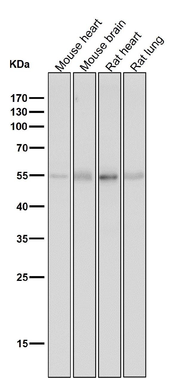
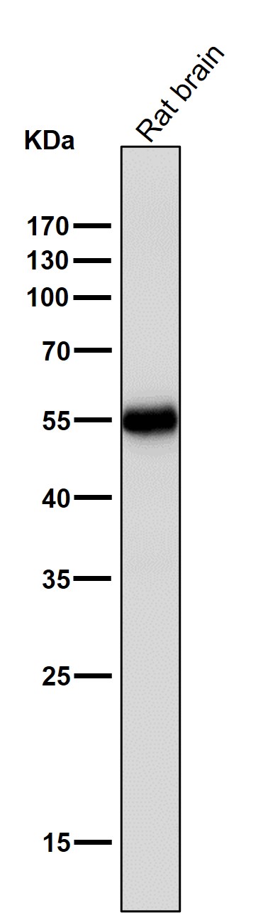
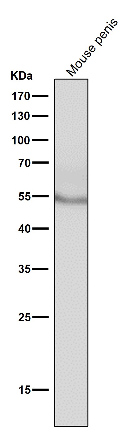
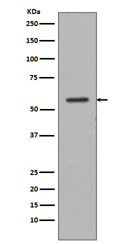
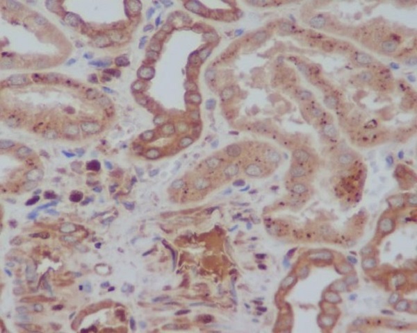
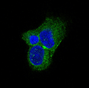
| Description | Anti-Tyrosine Hydroxylase TH Rabbit Monoclonal Antibody . Tested in WB, ICC/IF, Flow Cytometry applications. This antibody reacts with Human, Mouse, Rat. |
| Gene ID | 7054 |
|---|---|
| Other Names | Tyrosine 3-monooxygenase, 1.14.16.2 {ECO:0000269|PubMed:15287903, ECO:0000269|PubMed:1680128, ECO:0000269|PubMed:17391063, ECO:0000269|PubMed:24753243, ECO:0000269|PubMed:34922205, ECO:0000269|PubMed:8528210, ECO:0000269|Ref.18}, Tyrosine 3-hydroxylase, TH, TH (HGNC:11782), TYH |
| Calculated MW | 58600 MW KDa |
| Application Details | WB 1:500-1:2000 ICC/IF 1:50-1:200 FC 1:50 |
| Tissue Specificity | Mainly expressed in the brain and adrenal glands. |
| Contents | Rabbit IgG in phosphate buffered saline, pH 7.4, 150mM NaCl, 0.02% sodium azide and 50% glycerol, 0.4-0.5mg/ml BSA. |
| Clone Names | Clone: ECA-20 |
| Immunogen | A synthesized peptide derived from human Tyrosine Hydroxylase |
| Purification | Affinity-chromatography |
| Storage | Store at -20°C long term. Avoid freeze / thaw cycle. |
| Name | TH (HGNC:11782) |
|---|---|
| Synonyms | TYH |
| Function | Catalyzes the conversion of L-tyrosine to L- dihydroxyphenylalanine (L-Dopa), the rate-limiting step in the biosynthesis of catecholamines, dopamine, noradrenaline, and adrenaline. Uses tetrahydrobiopterin and molecular oxygen to convert tyrosine to L-Dopa (PubMed:15287903, PubMed:1680128, PubMed:17391063, PubMed:24753243, PubMed:34922205, PubMed:8528210, Ref.18). In addition to tyrosine, is able to catalyze the hydroxylation of phenylalanine and tryptophan with lower specificity (By similarity). Positively regulates the regression of retinal hyaloid vessels during postnatal development (By similarity). |
| Cellular Location | Cytoplasm, perinuclear region {ECO:0000250|UniProtKB:P24529}. Nucleus {ECO:0000250|UniProtKB:P04177} Cell projection, axon {ECO:0000250|UniProtKB:P24529}. Cytoplasm {ECO:0000250|UniProtKB:P04177}. Cytoplasmic vesicle, secretory vesicle, synaptic vesicle {ECO:0000250|UniProtKB:P04177}. Note=When phosphorylated at Ser-19 shows a nuclear distribution and when phosphorylated at Ser-31 as well at Ser-40 shows a cytosolic distribution (By similarity). Expressed in dopaminergic axons and axon terminals. {ECO:0000250|UniProtKB:P04177} |
| Tissue Location | Mainly expressed in the brain and adrenal glands. |
Research Areas
Citations (0)

Thousands of laboratories across the world have published research that depended on the performance of antibodies from Abcepta to advance their research. Check out links to articles that cite our products in major peer-reviewed journals, organized by research category.
Submit your citation using an Abcepta antibody to
info@abcepta.com, and receive a free "I Love Antibodies" mug.
info@abcepta.com, and receive a free "I Love Antibodies" mug.

Application Protocols
Provided below are standard protocols that you may find useful for product applications.
Abcepta welcomes feedback from its customers.
If you have used an Abcepta product and would like to share how it has performed, please click on the "Submit Review" button and provide the requested information. Our staff will examine and post your review and contact you if needed.
If you have any additional inquiries please email technical services at tech@abcepta.com.
$ 370.00
Cat# ABO14273
Ordering Information
United States
AlbaniaAustraliaAustriaBelgiumBosnia & HerzegovinaBrazilBulgariaCanadaCentral AmericaChinaCroatiaCyprusCzech RepublicDenmarkEstoniaFinlandFranceGermanyGreeceHong KongHungaryIcelandIndiaIndonesiaIrelandIsraelItalyJapanLatviaLithuaniaLuxembourgMacedoniaMalaysiaMaltaMexicoNetherlandsNew ZealandNorwayPakistanPolandPortugalRomaniaSerbiaSingaporeSlovakiaSloveniaSouth AfricaSouth KoreaSpainSwedenSwitzerlandTaiwanTurkeyUnited KingdomUnited StatesVietnamWorldwideOthers
USA Headquarters
(888) 735-7227 / (858) 622-0099 or (858) 875-1900
Other Products
Shipping Information
Domestic orders (in stock items)
Shipped out the same day. Orders placed after 1 PM (PST) will ship out the next business day.
International orders
Contact your local distributors
