


 Foundational characteristics of cancer include proliferation, angiogenesis, migration, evasion of apoptosis, and cellular immortality. Find key markers for these cellular processes and antibodies to detect them.
 The SUMOplot™ Analysis Program predicts and scores sumoylation sites in your protein. SUMOylation is a post-translational modification involved in various cellular processes, such as nuclear-cytosolic transport, transcriptional regulation, apoptosis, protein stability, response to stress, and progression through the cell cycle.
 The Autophagy Receptor Motif Plotter predicts and scores autophagy receptor binding sites in your protein. Identifying proteins connected to this pathway is critical to understanding the role of autophagy in physiological as well as pathological processes such as development, differentiation, neurodegenerative diseases, stress, infection, and cancer.
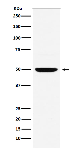
Anti-MUM1 Monoclonal Antibody
- SPECIFICATION
- CITATIONS
- PROTOCOLS
- BACKGROUND

Application
| WB, IHC, IF, ICC, IP, FC |
|---|---|
| Primary Accession | Q15306 |
| Host | Rabbit |
| Isotype | Rabbit IgG |
| Reactivity | Human |
| Clonality | Monoclonal |
| Format | Liquid |
| Description | Anti-MUM1 Monoclonal Antibody . Tested in WB, IHC, ICC/IF, IP, Flow Cytometry applications. This antibody reacts with Human. |
| Gene ID | 3662 |
|---|---|
| Other Names | Interferon regulatory factor 4, IRF-4, Lymphocyte-specific interferon regulatory factor, LSIRF, Multiple myeloma oncogene 1, NF-EM5, IRF4, MUM1 |
| Calculated MW | 51772 Da |
| Application Details | WB 1:1000-1:5000 IHC 1:50-1:200 ICC/IF 1:50-1:200 IP 1:100 FC 1:120 |
| Contents | Rabbit IgG in phosphate buffered saline, pH 7.4, 150mM NaCl, 0.02% sodium azide and 50% glycerol, 0.4-0.5mg/ml BSA. |
| Clone Names | Clone: ADOF-9 |
| Immunogen | A synthesized peptide derived from human MUM1 Melanoma associated antigen (mutated) 1 (MUM1, EXPAND1) is a PWWP-domain containing chromatin binding protein involved in maintaining chromatin architecture of interphase chromosomes. |
| Purification | Affinity-chromatography |
| Storage | Store at -20°C for one year. For short term storage and frequent use, store at 4°C for up to one month. Avoid repeated freeze-thaw cycles. |
| Name | IRF4 {ECO:0000303|PubMed:15489334, ECO:0000303|PubMed:8921401} |
|---|---|
| Function | Transcriptional activator. Binds to the interferon-stimulated response element (ISRE) of the MHC class I promoter. Binds the immunoglobulin lambda light chain enhancer, together with PU.1. Probably plays a role in ISRE-targeted signal transduction mechanisms specific to lymphoid cells. Involved in CD8(+) dendritic cell differentiation by forming a complex with the BATF-JUNB heterodimer in immune cells, leading to recognition of AICE sequence (5'-TGAnTCA/GAAA- 3'), an immune-specific regulatory element, followed by cooperative binding of BATF and IRF4 and activation of genes. |
| Cellular Location | Nucleus. Cytoplasm |
| Tissue Location | Lymphoid cells. |
Research Areas
Citations (0)

Thousands of laboratories across the world have published research that depended on the performance of antibodies from Abcepta to advance their research. Check out links to articles that cite our products in major peer-reviewed journals, organized by research category.
Submit your citation using an Abcepta antibody to
info@abcepta.com, and receive a free "I Love Antibodies" mug.
info@abcepta.com, and receive a free "I Love Antibodies" mug.

Application Protocols
Provided below are standard protocols that you may find useful for product applications.
Abcepta welcomes feedback from its customers.
If you have used an Abcepta product and would like to share how it has performed, please click on the "Submit Review" button and provide the requested information. Our staff will examine and post your review and contact you if needed.
If you have any additional inquiries please email technical services at tech@abcepta.com.
$ 370.00
Cat# ABO14493
Ordering Information
United States
AlbaniaAustraliaAustriaBelgiumBosnia & HerzegovinaBrazilBulgariaCanadaCentral AmericaChinaCroatiaCyprusCzech RepublicDenmarkEstoniaFinlandFranceGermanyGreeceHong KongHungaryIcelandIndiaIndonesiaIrelandIsraelItalyJapanLatviaLithuaniaLuxembourgMacedoniaMalaysiaMaltaMexicoNetherlandsNew ZealandNorwayPakistanPolandPortugalRomaniaSerbiaSingaporeSlovakiaSloveniaSouth AfricaSouth KoreaSpainSwedenSwitzerlandTaiwanTurkeyUnited KingdomUnited StatesVietnamWorldwideOthers
USA Headquarters
(888) 735-7227 / (858) 622-0099 or (858) 875-1900
Other Products
Shipping Information
Domestic orders (in stock items)
Shipped out the same day. Orders placed after 1 PM (PST) will ship out the next business day.
International orders
Contact your local distributors
