


 Foundational characteristics of cancer include proliferation, angiogenesis, migration, evasion of apoptosis, and cellular immortality. Find key markers for these cellular processes and antibodies to detect them.
 The SUMOplot™ Analysis Program predicts and scores sumoylation sites in your protein. SUMOylation is a post-translational modification involved in various cellular processes, such as nuclear-cytosolic transport, transcriptional regulation, apoptosis, protein stability, response to stress, and progression through the cell cycle.
 The Autophagy Receptor Motif Plotter predicts and scores autophagy receptor binding sites in your protein. Identifying proteins connected to this pathway is critical to understanding the role of autophagy in physiological as well as pathological processes such as development, differentiation, neurodegenerative diseases, stress, infection, and cancer.
> home > Products > Primary Antibodies > Antibody Collections > Head neck negative > Anti-Nogo Monoclonal Antibody
Anti-Nogo Monoclonal Antibody
- SPECIFICATION
- CITATIONS
- PROTOCOLS
- BACKGROUND

Application
| WB, IHC, IF, ICC, IP, FC |
|---|---|
| Primary Accession | Q9NQC3 |
| Host | Rabbit |
| Isotype | Rabbit IgG |
| Reactivity | Human |
| Clonality | Monoclonal |
| Format | Liquid |
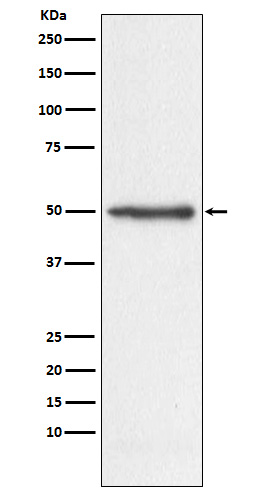
| Description | Anti-Nogo Monoclonal Antibody . Tested in WB, IHC, ICC/IF, IP, Flow Cytometry applications. This antibody reacts with Human. |
| Gene ID | 57142 |
|---|---|
| Other Names | Reticulon-4, Foocen, Neurite outgrowth inhibitor, Nogo protein, Neuroendocrine-specific protein, NSP, Neuroendocrine-specific protein C homolog, RTN-x, Reticulon-5, RTN4 (HGNC:14085) |
| Calculated MW | 129931 Da |
| Application Details | WB 1:500-1:2000 IHC 1:50-1:200 ICC/IF 1:50-1:200 IP 1:50 FC 1:50 |
| Contents | Rabbit IgG in phosphate buffered saline, pH 7.4, 150mM NaCl, 0.02% sodium azide and 50% glycerol, 0.4-0.5mg/ml BSA. |
| Clone Names | Clone: AEAH-18 |
| Immunogen | A synthesized peptide derived from human Nogo Developmental neurite growth regulatory factor with a role as a negative regulator of axon-axon adhesion and growth, and as a facilitator of neurite branching. Regulates neurite fasciculation, branching and extension in the developing nervous system. |
| Purification | Affinity-chromatography |
| Storage | Store at -20°C for one year. For short term storage and frequent use, store at 4°C for up to one month. Avoid repeated freeze-thaw cycles. |
| Name | RTN4 (HGNC:14085) |
|---|---|
| Function | Required to induce the formation and stabilization of endoplasmic reticulum (ER) tubules (PubMed:24262037, PubMed:25612671, PubMed:27619977). They regulate membrane morphogenesis in the ER by promoting tubular ER production (PubMed:24262037, PubMed:25612671, PubMed:27619977, PubMed:27786289). They influence nuclear envelope expansion, nuclear pore complex formation and proper localization of inner nuclear membrane proteins (PubMed:26906412). However each isoform have specific functions mainly depending on their tissue expression specificities (Probable). |
| Cellular Location | [Isoform A]: Endoplasmic reticulum membrane; Multi-pass membrane protein. Cell membrane; Multi-pass membrane protein; Cytoplasmic side Synapse {ECO:0000250|UniProtKB:Q99P72}. Note=Anchored to the membrane of the endoplasmic reticulum (ER) through 2 putative transmembrane domains. Localizes throughout the ER tubular network (PubMed:27619977) Co-localizes with TMEM33 at the ER sheets [Isoform C]: Endoplasmic reticulum membrane; Multi-pass membrane protein |
| Tissue Location | Isoform A: is specifically expressed in brain and testis and weakly in heart and skeletal muscle. Isoform B: widely expressed except for the liver. Highly expressed in endothelial cells and vascular smooth muscle cells, including blood vessels and mesenteric arteries (PubMed:15034570, PubMed:21183689). Isoform C: is expressed in brain, skeletal muscle and adipocytes. Isoform D is testis-specific. |
Research Areas
Citations (0)

Thousands of laboratories across the world have published research that depended on the performance of antibodies from Abcepta to advance their research. Check out links to articles that cite our products in major peer-reviewed journals, organized by research category.
Submit your citation using an Abcepta antibody to
info@abcepta.com, and receive a free "I Love Antibodies" mug.
info@abcepta.com, and receive a free "I Love Antibodies" mug.

Application Protocols
Provided below are standard protocols that you may find useful for product applications.
Abcepta welcomes feedback from its customers.
If you have used an Abcepta product and would like to share how it has performed, please click on the "Submit Review" button and provide the requested information. Our staff will examine and post your review and contact you if needed.
If you have any additional inquiries please email technical services at tech@abcepta.com.
$ 370.00
Cat# ABO14605
Ordering Information
United States
AlbaniaAustraliaAustriaBelgiumBosnia & HerzegovinaBrazilBulgariaCanadaCentral AmericaChinaCroatiaCyprusCzech RepublicDenmarkEstoniaFinlandFranceGermanyGreeceHong KongHungaryIcelandIndiaIndonesiaIrelandIsraelItalyJapanLatviaLithuaniaLuxembourgMacedoniaMalaysiaMaltaMexicoNetherlandsNew ZealandNorwayPakistanPolandPortugalRomaniaSerbiaSingaporeSlovakiaSloveniaSouth AfricaSouth KoreaSpainSwedenSwitzerlandTaiwanTurkeyUnited KingdomUnited StatesVietnamWorldwideOthers
USA Headquarters
(888) 735-7227 / (858) 622-0099 or (858) 875-1900
Other Products
Shipping Information
Domestic orders (in stock items)
Shipped out the same day. Orders placed after 1 PM (PST) will ship out the next business day.
International orders
Contact your local distributors
