


 Foundational characteristics of cancer include proliferation, angiogenesis, migration, evasion of apoptosis, and cellular immortality. Find key markers for these cellular processes and antibodies to detect them.
 The SUMOplot™ Analysis Program predicts and scores sumoylation sites in your protein. SUMOylation is a post-translational modification involved in various cellular processes, such as nuclear-cytosolic transport, transcriptional regulation, apoptosis, protein stability, response to stress, and progression through the cell cycle.
 The Autophagy Receptor Motif Plotter predicts and scores autophagy receptor binding sites in your protein. Identifying proteins connected to this pathway is critical to understanding the role of autophagy in physiological as well as pathological processes such as development, differentiation, neurodegenerative diseases, stress, infection, and cancer.
Anti-RBPJK Monoclonal Antibody
- SPECIFICATION
- CITATIONS
- PROTOCOLS
- BACKGROUND

Application
| WB, IF, ICC |
|---|---|
| Primary Accession | Q06330 |
| Host | Rabbit |
| Isotype | Rabbit IgG |
| Reactivity | Rat, Human, Mouse |
| Clonality | Monoclonal |
| Format | Liquid |
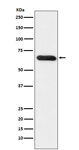
| Description | Anti-RBPJK Monoclonal Antibody . Tested in WB, ICC/IF applications. This antibody reacts with Human, Mouse, Rat. |
| Gene ID | 3516 |
|---|---|
| Other Names | Recombining binding protein suppressor of hairless, CBF-1, J kappa-recombination signal-binding protein, RBP-J kappa, RBP-J, RBP-JK, Renal carcinoma antigen NY-REN-30, RBPJ (HGNC:5724) |
| Calculated MW | 55637 Da |
| Application Details | WB 1:500-1:2000 ICC/IF 1:50-1:200 |
| Contents | Rabbit IgG in phosphate buffered saline, pH 7.4, 150mM NaCl, 0.02% sodium azide and 50% glycerol, 0.4-0.5mg/ml BSA. |
| Clone Names | Clone: AEEF-18 |
| Immunogen | A synthesized peptide derived from human RBPJK Transcriptional regulator that plays a central role in Notch signaling, a signaling pathway involved in cell-cell communication that regulates a broad spectrum of cell-fate determinations. Acts as a transcriptional repressor when it is not associated with Notch proteins. |
| Purification | Affinity-chromatography |
| Storage | Store at -20°C for one year. For short term storage and frequent use, store at 4°C for up to one month. Avoid repeated freeze-thaw cycles. |
| Name | RBPJ (HGNC:5724) |
|---|---|
| Function | Transcriptional regulator that plays a central role in Notch signaling, a signaling pathway involved in cell-cell communication that regulates a broad spectrum of cell-fate determinations. Acts as a transcriptional repressor when it is not associated with Notch proteins. When associated with some NICD product of Notch proteins (Notch intracellular domain), it acts as a transcriptional activator that activates transcription of Notch target genes. Probably represses or activates transcription via the recruitment of chromatin remodeling complexes containing histone deacetylase or histone acetylase proteins, respectively. Specifically binds to the immunoglobulin kappa-type J segment recombination signal sequence. Binds specifically to methylated DNA (PubMed:21991380). Binds to the oxygen responsive element of COX4I2 and activates its transcription under hypoxia conditions (4% oxygen) (PubMed:23303788). Negatively regulates the phagocyte oxidative burst in response to bacterial infection by repressing transcription of NADPH oxidase subunits (By similarity). |
| Cellular Location | Nucleus. Cytoplasm. Note=Mainly nuclear, upon interaction with RITA/C12orf52, translocates to the cytoplasm, down- regulating the Notch signaling pathway |
Research Areas
Citations (0)

Thousands of laboratories across the world have published research that depended on the performance of antibodies from Abcepta to advance their research. Check out links to articles that cite our products in major peer-reviewed journals, organized by research category.
Submit your citation using an Abcepta antibody to
info@abcepta.com, and receive a free "I Love Antibodies" mug.
info@abcepta.com, and receive a free "I Love Antibodies" mug.

Application Protocols
Provided below are standard protocols that you may find useful for product applications.
Abcepta welcomes feedback from its customers.
If you have used an Abcepta product and would like to share how it has performed, please click on the "Submit Review" button and provide the requested information. Our staff will examine and post your review and contact you if needed.
If you have any additional inquiries please email technical services at tech@abcepta.com.
$ 370.00
Cat# ABO14643
Ordering Information
United States
AlbaniaAustraliaAustriaBelgiumBosnia & HerzegovinaBrazilBulgariaCanadaCentral AmericaChinaCroatiaCyprusCzech RepublicDenmarkEstoniaFinlandFranceGermanyGreeceHong KongHungaryIcelandIndiaIndonesiaIrelandIsraelItalyJapanLatviaLithuaniaLuxembourgMacedoniaMalaysiaMaltaMexicoNetherlandsNew ZealandNorwayPakistanPolandPortugalRomaniaSerbiaSingaporeSlovakiaSloveniaSouth AfricaSouth KoreaSpainSwedenSwitzerlandTaiwanTurkeyUnited KingdomUnited StatesVietnamWorldwideOthers
USA Headquarters
(888) 735-7227 / (858) 622-0099 or (858) 875-1900
Other Products
Shipping Information
Domestic orders (in stock items)
Shipped out the same day. Orders placed after 1 PM (PST) will ship out the next business day.
International orders
Contact your local distributors
