


 Foundational characteristics of cancer include proliferation, angiogenesis, migration, evasion of apoptosis, and cellular immortality. Find key markers for these cellular processes and antibodies to detect them.
 The SUMOplot™ Analysis Program predicts and scores sumoylation sites in your protein. SUMOylation is a post-translational modification involved in various cellular processes, such as nuclear-cytosolic transport, transcriptional regulation, apoptosis, protein stability, response to stress, and progression through the cell cycle.
 The Autophagy Receptor Motif Plotter predicts and scores autophagy receptor binding sites in your protein. Identifying proteins connected to this pathway is critical to understanding the role of autophagy in physiological as well as pathological processes such as development, differentiation, neurodegenerative diseases, stress, infection, and cancer.
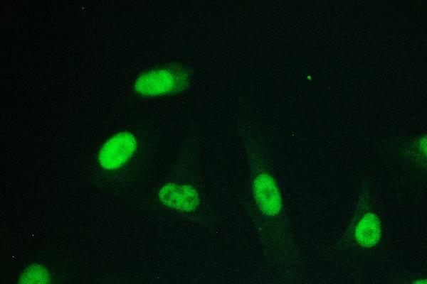
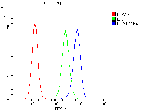
Anti-RPA70 RPA1 Antibody Picoband™ (monoclonal, 11H4)
- SPECIFICATION
- CITATIONS
- PROTOCOLS
- BACKGROUND

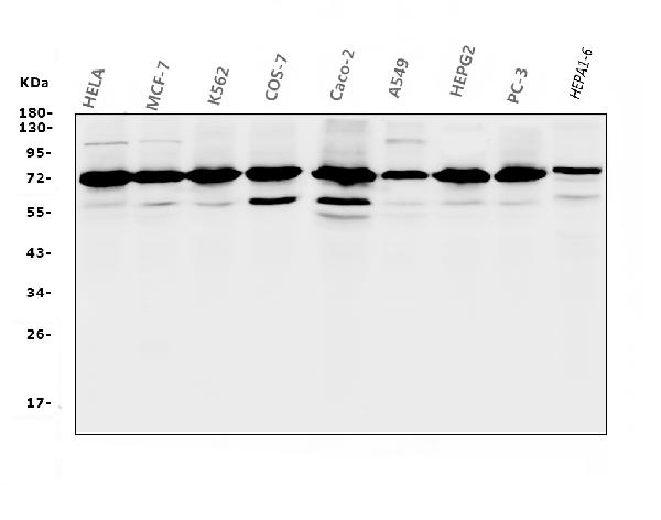
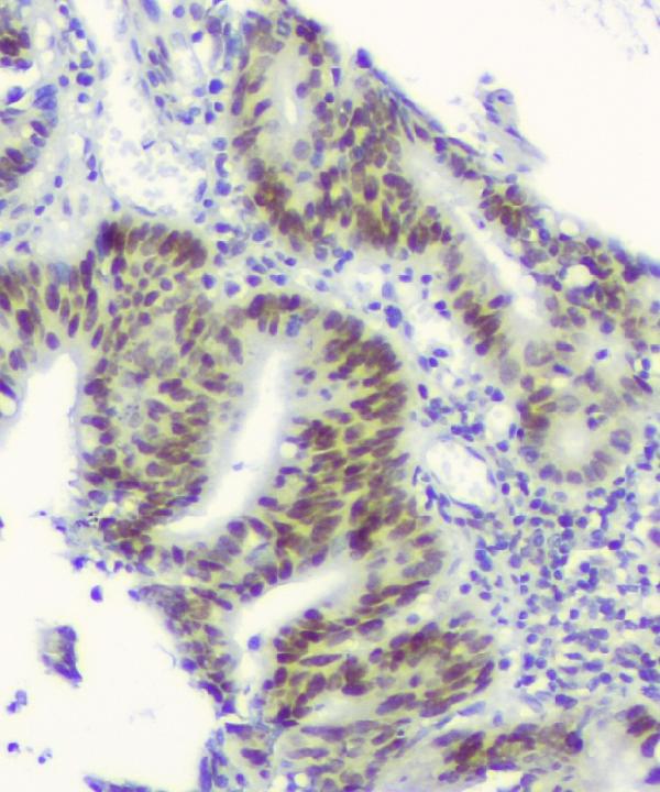
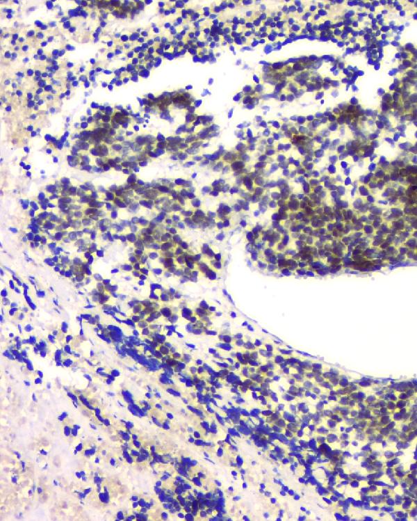
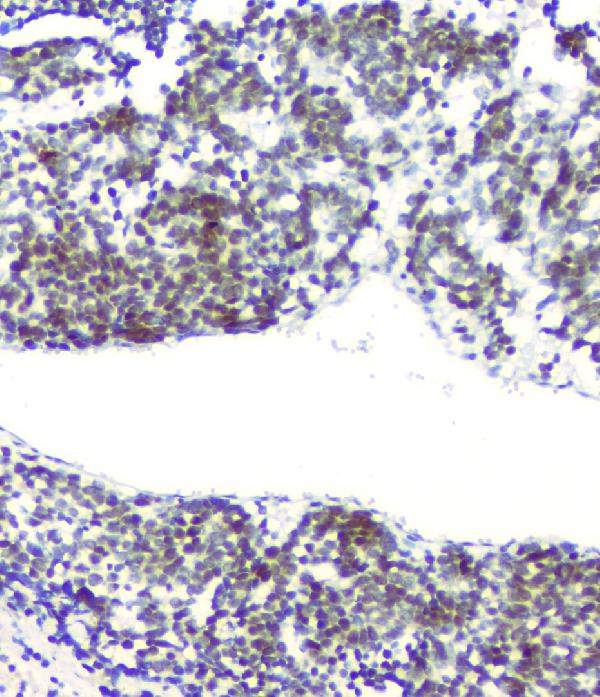
Application
| WB, IHC, IF, FC |
|---|---|
| Primary Accession | P27694 |
| Host | Mouse |
| Isotype | Mouse IgG2b |
| Reactivity | Human, Mouse, Monkey |
| Clonality | Monoclonal |
| Format | Lyophilized |
| Description | Anti-RPA70 RPA1 Antibody Picoband™ (monoclonal, 11H4) . Tested in Flow Cytometry, IF, IHC, WB applications. This antibody reacts with Human, Monkey, Mouse. |
| Gene ID | 6117 |
|---|---|
| Other Names | Replication protein A 70 kDa DNA-binding subunit, RP-A p70, Replication factor A protein 1, RF-A protein 1, Single-stranded DNA-binding protein, Replication protein A 70 kDa DNA-binding subunit, N-terminally processed, RPA1, REPA1, RPA70 |
| Calculated MW | 12 kDa |
| Application Details | Western blot, 0.1-0.5 µg/ml Immunohistochemistry (Paraffin-embedded Section), 0.5-1 µg/ml Immunofluorescence, 2 µg/ml Flow Cytometry, 1-3 µg/1x10^6 cells |
| Subcellular Localization | Nucleus |
| Contents | Each vial contains 4mg Trehalose, 0.9mg NaCl, 0.2mg Na2HPO4, 0.05mg NaN3. |
| Clone Names | Clone: 11H4 |
| Immunogen | A synthetic peptide corresponding to a sequence at the C-terminus of human RPA70, different from the related mouse sequence by three amino acids. |
| Cross Reactivity | No cross-reactivity with other proteins. |
| Storage | Store at -20˚C for one year from date of receipt. After reconstitution, at 4˚C for one month. It can also be aliquotted and stored frozen at -20˚C for six months. Avoid repeated freeze-thaw cycles. |
| Name | RPA1 |
|---|---|
| Synonyms | REPA1, RPA70 |
| Function | As part of the heterotrimeric replication protein A complex (RPA/RP-A), binds and stabilizes single-stranded DNA intermediates that form during DNA replication or upon DNA stress. It prevents their reannealing and in parallel, recruits and activates different proteins and complexes involved in DNA metabolism (PubMed:17596542, PubMed:27723717, PubMed:27723720). Thereby, it plays an essential role both in DNA replication and the cellular response to DNA damage (PubMed:9430682). In the cellular response to DNA damage, the RPA complex controls DNA repair and DNA damage checkpoint activation. Through recruitment of ATRIP activates the ATR kinase a master regulator of the DNA damage response (PubMed:24332808). It is required for the recruitment of the DNA double-strand break repair factors RAD51 and RAD52 to chromatin in response to DNA damage (PubMed:17765923). Also recruits to sites of DNA damage proteins like XPA and XPG that are involved in nucleotide excision repair and is required for this mechanism of DNA repair (PubMed:7697716). Also plays a role in base excision repair (BER) probably through interaction with UNG (PubMed:9765279). Also recruits SMARCAL1/HARP, which is involved in replication fork restart, to sites of DNA damage. Plays a role in telomere maintenance (PubMed:17959650, PubMed:34767620). As part of the alternative replication protein A complex, aRPA, binds single-stranded DNA and probably plays a role in DNA repair. Compared to the RPA2- containing, canonical RPA complex, may not support chromosomal DNA replication and cell cycle progression through S-phase. The aRPA may not promote efficient priming by DNA polymerase alpha but could support DNA synthesis by polymerase delta in presence of PCNA and replication factor C (RFC), the dual incision/excision reaction of nucleotide excision repair and RAD51-dependent strand exchange (PubMed:19996105). RPA stimulates 5'-3' helicase activity of the BRIP1/FANCJ (PubMed:17596542). |
| Cellular Location | Nucleus. Nucleus, PML body. Note=Enriched in PML bodies in cells displaying alternative lengthening of their telomeres |

Thousands of laboratories across the world have published research that depended on the performance of antibodies from Abcepta to advance their research. Check out links to articles that cite our products in major peer-reviewed journals, organized by research category.
info@abcepta.com, and receive a free "I Love Antibodies" mug.

Provided below are standard protocols that you may find useful for product applications.
Background
Replication protein A 70 kDa DNA-binding subunit is a protein that in humans is encoded by the RPA1 gene. This gene is mapped to chromosome 17p13.3. Replication protein A (RPA) is a heterotrimeric single-strand DNA (ssDNA)-binding protein essential for DNA replication, repair, and recombination. It is composed of 70-kD (RPA1), 32-kD (RPA2), and 14-kD (RPA3) subunits. The RPA1 subunit is responsible for high-affinity ssDNA binding. The RPA complex was originally isolated as a factor essential for in vitro replication of the papovavirus SV40. It had been found that recombinant human RPA1, purified from bacteria, exhibited ssDNA-binding activity comparable to that of the complete RPA complex. RPA1 could substitute for the complete complex in stimulating the activity of DNA polymerase alpha-primase, but it could not substitute for the complete complex in SV40 DNA replication in vitro, suggesting an important functional role for the other subunits.
If you have used an Abcepta product and would like to share how it has performed, please click on the "Submit Review" button and provide the requested information. Our staff will examine and post your review and contact you if needed.
If you have any additional inquiries please email technical services at tech@abcepta.com.
Ordering Information
Other Products
Shipping Information
