


 Foundational characteristics of cancer include proliferation, angiogenesis, migration, evasion of apoptosis, and cellular immortality. Find key markers for these cellular processes and antibodies to detect them.
 The SUMOplot™ Analysis Program predicts and scores sumoylation sites in your protein. SUMOylation is a post-translational modification involved in various cellular processes, such as nuclear-cytosolic transport, transcriptional regulation, apoptosis, protein stability, response to stress, and progression through the cell cycle.
 The Autophagy Receptor Motif Plotter predicts and scores autophagy receptor binding sites in your protein. Identifying proteins connected to this pathway is critical to understanding the role of autophagy in physiological as well as pathological processes such as development, differentiation, neurodegenerative diseases, stress, infection, and cancer.
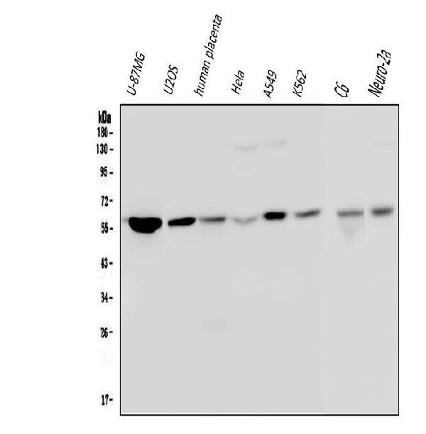
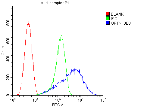
Anti-Optineurin/OPTN Antibody Picoband™ (monoclonal, 3D8)
- SPECIFICATION
- CITATIONS
- PROTOCOLS
- BACKGROUND

Application
| WB, FC |
|---|---|
| Primary Accession | Q96CV9 |
| Host | Mouse |
| Isotype | Mouse IgG2b |
| Reactivity | Rat, Human, Mouse |
| Clonality | Monoclonal |
| Format | Lyophilized |
| Description | Anti-Optineurin/OPTN Antibody Picoband™ (monoclonal, 3D8) . Tested in Flow Cytometry, WB applications. This antibody reacts with Human, Mouse, Rat. |
| Reconstitution | Add 0.2ml of distilled water will yield a concentration of 500 µg/ml. |
| Gene ID | 10133 |
|---|---|
| Other Names | Optineurin, E3-14.7K-interacting protein, FIP-2, Huntingtin yeast partner L, Huntingtin-interacting protein 7, HIP-7, Huntingtin-interacting protein L, NEMO-related protein, Optic neuropathy-inducing protein, Transcription factor IIIA-interacting protein, TFIIIA-IntP, OPTN |
| Calculated MW | 66 kDa |
| Application Details | Western blot, 0.1-0.5 µg/ml Flow Cytometry, 1-3 µg/1x10^6 cells |
| Subcellular Localization | Golgi apparatus. Trans-Golgi network. Recycling endosome. Perinuclear region. Autophagosome. Cytoplasmic vesicle. |
| Tissue Specificity | Present in aqueous humor of the eye. Highly expressed in trabecular meshwork. Expressed nonpigmented ciliary epithelium, retina, brain, adrenal cortex, fetus, lymphocyte, fibroblast, skeletal muscle, heart, liver, brain and placenta. |
| Contents | Each vial contains 4mg Trehalose, 0.9mg NaCl, 0.2mg Na2HPO4, 0.05mg NaN3. |
| Clone Names | Clone: 3D8 |
| Immunogen | E.coli-derived human Optineurin recombinant protein (Position: R241-I577). Human Optineurin shares 82% amino acid (aa) sequence identity with both mouse and rat Optineurin. |
| Cross Reactivity | No cross-reactivity with other proteins. |
| Storage | Store at -20˚C for one year from date of receipt. After reconstitution, at 4˚C for one month. It can also be aliquotted and stored frozen at -20˚C for six months. Avoid repeated freeze-thaw cycles. |
| Name | OPTN |
|---|---|
| Function | Plays an important role in the maintenance of the Golgi complex, in membrane trafficking, in exocytosis, through its interaction with myosin VI and Rab8 (PubMed:27534431). Links myosin VI to the Golgi complex and plays an important role in Golgi ribbon formation (PubMed:27534431). Plays a role in the activation of innate immune response during viral infection. Mechanistically, recruits TBK1 at the Golgi apparatus, promoting its trans-phosphorylation after RLR or TLR3 stimulation (PubMed:27538435). In turn, activated TBK1 phosphorylates its downstream partner IRF3 to produce IFN-beta/IFNB1. Plays a neuroprotective role in the eye and optic nerve. May act by regulating membrane trafficking and cellular morphogenesis via a complex that contains Rab8 and huntingtin (HD). Mediates the interaction of Rab8 with the probable GTPase-activating protein TBC1D17 during Rab8-mediated endocytic trafficking, such as that of transferrin receptor (TFRC/TfR); regulates Rab8 recruitment to tubules emanating from the endocytic recycling compartment (PubMed:22854040). Autophagy receptor that interacts directly with both the cargo to become degraded and an autophagy modifier of the MAP1 LC3 family; targets ubiquitin- coated bacteria (xenophagy), such as cytoplasmic Salmonella enterica, and appears to function in the same pathway as SQSTM1 and CALCOCO2/NDP52. |
| Cellular Location | Cytoplasm, perinuclear region. Golgi apparatus. Golgi apparatus, trans-Golgi network Cytoplasmic vesicle, autophagosome. Cytoplasmic vesicle. Recycling endosome. Note=Found in the perinuclear region and associates with the Golgi apparatus (PubMed:27534431) Colocalizes with MYO6 and RAB8 at the Golgi complex and in vesicular structures close to the plasma membrane. Localizes to LC3-positive cytoplasmic vesicles upon induction of autophagy |
| Tissue Location | Present in aqueous humor of the eye (at protein level). Expressed in the trabecular meshwork (at protein level) (PubMed:11834836, PubMed:12379221, PubMed:12646749). Expressed in nonpigmented ciliary epithelium (at protein level) (PubMed:11834836) Expressed at high levels in skeletal muscle, also detected in heart, brain, pancreas, kidney, placenta and liver (PubMed:9488477). Expressed in dermal fibroblasts (at protein level) (PubMed:11834836) |

Thousands of laboratories across the world have published research that depended on the performance of antibodies from Abcepta to advance their research. Check out links to articles that cite our products in major peer-reviewed journals, organized by research category.
info@abcepta.com, and receive a free "I Love Antibodies" mug.

Provided below are standard protocols that you may find useful for product applications.
Background
OPTN is also known as NRP, FIP2 or HYPL. This gene encodes the coiled-coil containing protein optineurin. Optineurin may play a role in normal-tension glaucoma and adult-onset primary open angle glaucoma. Optineurin interacts with adenovirus E3-14.7K protein and may utilize tumor necrosis factor-alpha or Fas-ligand pathways to mediate apoptosis, inflammation or vasoconstriction. Optineurin may also function in cellular morphogenesis and membrane trafficking, vesicle trafficking, and transcription activation through its interactions with the RAB8, huntingtin, and transcription factor IIIA proteins. Alternative splicing results in multiple transcript variants encoding the same protein.
If you have used an Abcepta product and would like to share how it has performed, please click on the "Submit Review" button and provide the requested information. Our staff will examine and post your review and contact you if needed.
If you have any additional inquiries please email technical services at tech@abcepta.com.
Ordering Information
Other Products
Shipping Information
