


 Foundational characteristics of cancer include proliferation, angiogenesis, migration, evasion of apoptosis, and cellular immortality. Find key markers for these cellular processes and antibodies to detect them.
 The SUMOplot™ Analysis Program predicts and scores sumoylation sites in your protein. SUMOylation is a post-translational modification involved in various cellular processes, such as nuclear-cytosolic transport, transcriptional regulation, apoptosis, protein stability, response to stress, and progression through the cell cycle.
 The Autophagy Receptor Motif Plotter predicts and scores autophagy receptor binding sites in your protein. Identifying proteins connected to this pathway is critical to understanding the role of autophagy in physiological as well as pathological processes such as development, differentiation, neurodegenerative diseases, stress, infection, and cancer.
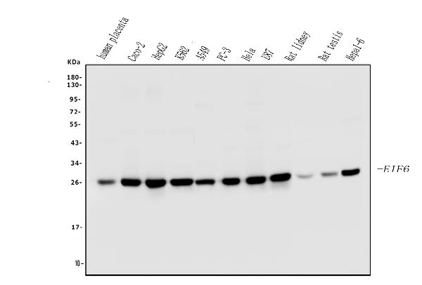
Anti-EIF6 Antibody Picoband™ (monoclonal, 2I11)
- SPECIFICATION
- CITATIONS
- PROTOCOLS
- BACKGROUND

Application
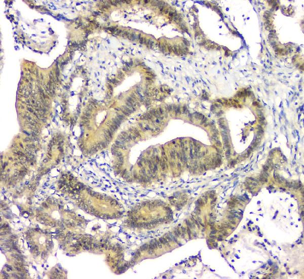
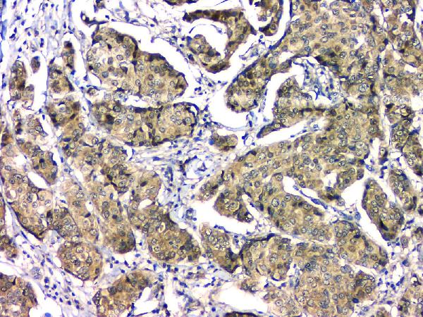
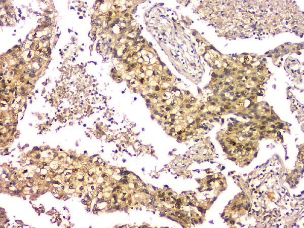
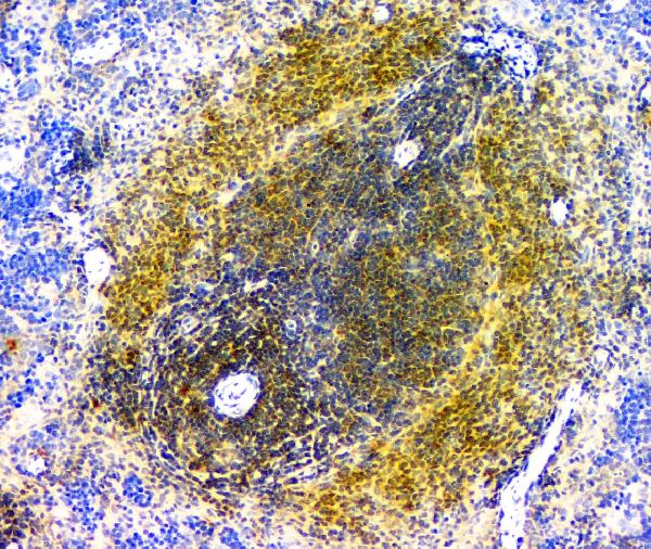
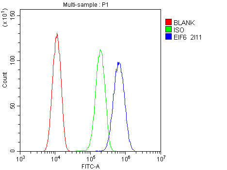
| WB, IHC, FC |
|---|---|
| Primary Accession | P56537 |
| Host | Mouse |
| Isotype | Mouse IgG2a |
| Reactivity | Rat, Human, Mouse |
| Clonality | Monoclonal |
| Format | Lyophilized |
| Description | Anti-EIF6 Antibody Picoband™ (monoclonal, 2I11) . Tested in Flow Cytometry, IHC, WB applications. This antibody reacts with Human, Mouse, Rat. |
| Reconstitution | Add 0.2ml of distilled water will yield a concentration of 500 µg/ml. |
| Gene ID | 3692 |
|---|---|
| Other Names | Eukaryotic translation initiation factor 6 {ECO:0000255|HAMAP-Rule:MF_03132}, eIF-6 {ECO:0000255|HAMAP-Rule:MF_03132}, B(2)GCN homolog, B4 integrin interactor, CAB, p27(BBP), EIF6 {ECO:0000255|HAMAP-Rule:MF_03132, ECO:0000312|HGNC:HGNC:6159} |
| Calculated MW | 26 kDa |
| Application Details | Western blot, 0.1-0.5 µg/ml Immunohistochemistry (Paraffin-embedded Section), 0.5-1 µg/ml Flow Cytometry, 1-3 µg/1x10^6 cells |
| Subcellular Localization | nucleolus. Cytoplasm |
| Tissue Specificity | Expressed at very high levels in colon carcinoma with lower levels in normal colon and ileum and lowest levels in kidney and muscle. |
| Contents | Each vial contains 4mg Trehalose, 0.9mg NaCl, 0.2mg Na2HPO4, 0.05mg NaN3. |
| Clone Names | Clone: 2I11 |
| Immunogen | E.coli-derived human EIF6 recombinant protein (Position: N66-T210). Human EIF6 shares 99.3% amino acid (aa) sequence identity with both mouse and rat EIF6. |
| Cross Reactivity | No cross-reactivity with other proteins. |
| Storage | Store at -20˚C for one year from date of receipt. After reconstitution, at 4˚C for one month. It can also be aliquotted and stored frozen at -20˚C for six months. Avoid repeated freeze-thaw cycles. |
| Name | EIF6 {ECO:0000255|HAMAP-Rule:MF_03132, ECO:0000312|HGNC:HGNC:6159} |
|---|---|
| Function | Binds to the 60S ribosomal subunit and prevents its association with the 40S ribosomal subunit to form the 80S initiation complex in the cytoplasm (PubMed:10085284, PubMed:14654845, PubMed:21536732, PubMed:32669547). Behaves as a stimulatory translation initiation factor downstream insulin/growth factors. Is also involved in ribosome biogenesis. Associates with pre-60S subunits in the nucleus and is involved in its nuclear export. Cytoplasmic release of TIF6 from 60S subunits and nuclear relocalization is promoted by a RACK1 (RACK1)- dependent protein kinase C activity (PubMed:10085284, PubMed:14654845, PubMed:21536732). In tissues responsive to insulin, controls fatty acid synthesis and glycolysis by exerting translational control of adipogenic transcription factors such as CEBPB, CEBPD and ATF4 that have G/C rich or uORF in their 5'UTR. Required for ROS-dependent megakaryocyte maturation and platelets formation, controls the expression of mitochondrial respiratory chain genes involved in reactive oxygen species (ROS) synthesis (By similarity). Involved in miRNA-mediated gene silencing by the RNA-induced silencing complex (RISC). Required for both miRNA-mediated translational repression and miRNA-mediated cleavage of complementary mRNAs by RISC (PubMed:17507929). Modulates cell cycle progression and global translation of pre-B cells, its activation seems to be rate-limiting in tumorigenesis and tumor growth (By similarity). |
| Cellular Location | Cytoplasm. Nucleus, nucleolus. Note=Shuttles between cytoplasm and nucleus/nucleolus |
| Tissue Location | Expressed at very high levels in colon carcinoma with lower levels in normal colon and ileum and lowest levels in kidney and muscle (at protein level). |

Thousands of laboratories across the world have published research that depended on the performance of antibodies from Abcepta to advance their research. Check out links to articles that cite our products in major peer-reviewed journals, organized by research category.
info@abcepta.com, and receive a free "I Love Antibodies" mug.

Provided below are standard protocols that you may find useful for product applications.
Background
EIF6 (Eukaryotic Translation Initiation Factor 6), also called EIF3A or ITGB4BP, is a human gene. By fluorescence in situ hybridization, Sanvito et al. (1998) mapped the ITGB4BP gene to 20q11.2. Ceci et al. (2003) demonstrated that the ribosomal 60S subunit is activated by release of EIF6. In the cytoplasm, EIF6 is bound to free 60S but not to 80S subunits. Furthermore, EIF6 interacts in the cytoplasm with RACK1, a receptor for activated protein kinase C. Gandin et al. (2008) demonstrated that mammalian eIF6 is required for efficient initiation of translation in vivo. Eif6-null mouse embryos were lethal at preimplantation. Heterozygous mice had 50% reduction of eIF6 levels in all tissues, and showed reduced mass of hepatic and adipose tissues due to a lower number of cells and to impaired G1/S cell cycle progression.
If you have used an Abcepta product and would like to share how it has performed, please click on the "Submit Review" button and provide the requested information. Our staff will examine and post your review and contact you if needed.
If you have any additional inquiries please email technical services at tech@abcepta.com.
Ordering Information
Other Products
Shipping Information
