


 Foundational characteristics of cancer include proliferation, angiogenesis, migration, evasion of apoptosis, and cellular immortality. Find key markers for these cellular processes and antibodies to detect them.
 The SUMOplot™ Analysis Program predicts and scores sumoylation sites in your protein. SUMOylation is a post-translational modification involved in various cellular processes, such as nuclear-cytosolic transport, transcriptional regulation, apoptosis, protein stability, response to stress, and progression through the cell cycle.
 The Autophagy Receptor Motif Plotter predicts and scores autophagy receptor binding sites in your protein. Identifying proteins connected to this pathway is critical to understanding the role of autophagy in physiological as well as pathological processes such as development, differentiation, neurodegenerative diseases, stress, infection, and cancer.
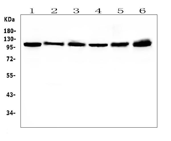
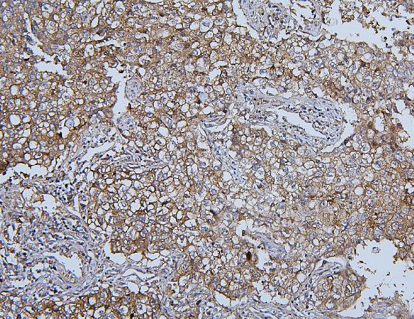
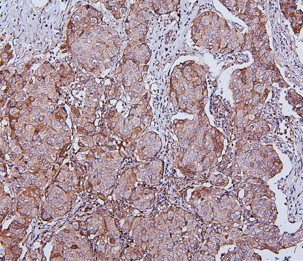
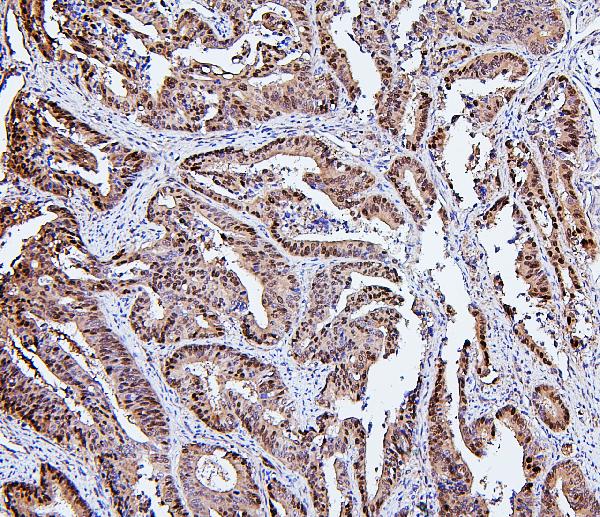
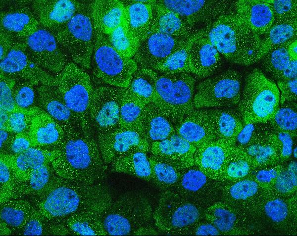
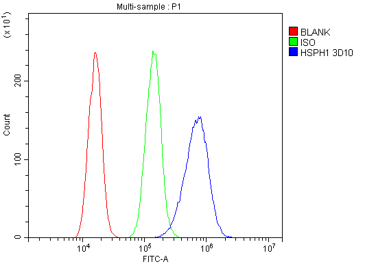
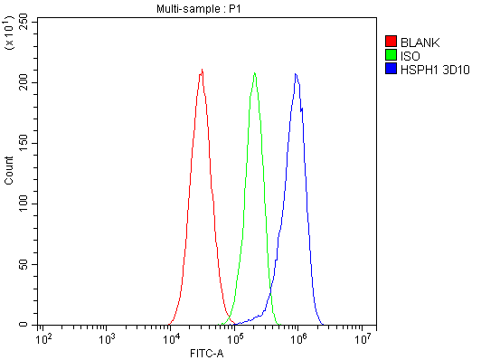
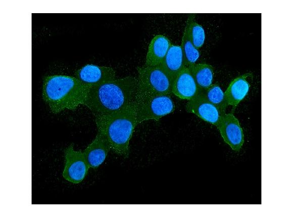
Anti-Hsp105/HSPH1 Antibody Picoband™ (monoclonal, 3D10)
- SPECIFICATION
- CITATIONS
- PROTOCOLS
- BACKGROUND

Application
| WB, IHC, IF, ICC, FC |
|---|---|
| Primary Accession | Q92598 |
| Host | Mouse |
| Isotype | Mouse IgG1 |
| Reactivity | Human |
| Clonality | Monoclonal |
| Format | Lyophilized |
| Description | Anti-Hsp105/HSPH1 Antibody Picoband™ (monoclonal, 3D10) . Tested in Flow Cytometry, IF, IHC, ICC, WB applications. This antibody reacts with Human. |
| Reconstitution | Add 0.2ml of distilled water will yield a concentration of 500 µg/ml. |
| Gene ID | 10808 |
|---|---|
| Other Names | Heat shock protein 105 kDa, Antigen NY-CO-25, Heat shock 110 kDa protein, Heat shock protein family H member 1, HSPH1, HSP105, HSP110, KIAA0201 |
| Calculated MW | 105 kDa |
| Application Details | Western blot, 0.1-0.5 µg/ml, Human Immunohistochemistry (Paraffin-embedded Section), 0.5-1 µg/ml, Human, By Heat Immunocytochemistry/Immunofluorescence, 2 µg/ml, Human Flow Cytometry, 1-3 µg/1x10^6 cells, Human |
| Subcellular Localization | Cytoplasm. |
| Tissue Specificity | Highly expressed in testis. Present at lower levels in most brain regions, except cerebellum. Overexpressed in cancer cells. |
| Contents | Each vial contains 4mg Trehalose, 0.9mg NaCl, 0.2mg Na2HPO4, 0.05mg NaN3. |
| Clone Names | Clone: 3D10 |
| Immunogen | E. coli-derived human Hsp105 recombinant protein (Position: Y653-D858). |
| Cross Reactivity | No cross-reactivity with other proteins. |
| Storage | Store at -20˚C for one year from date of receipt. After reconstitution, at 4˚C for one month. It can also be aliquotted and stored frozen at -20˚C for six months. Avoid repeated freeze-thaw cycles. |
| Name | HSPH1 |
|---|---|
| Synonyms | HSP105, HSP110, KIAA0201 |
| Function | Acts as a nucleotide-exchange factor (NEF) for chaperone proteins HSPA1A and HSPA1B, promoting the release of ADP from HSPA1A/B thereby triggering client/substrate protein release (PubMed:24318877). Prevents the aggregation of denatured proteins in cells under severe stress, on which the ATP levels decrease markedly. Inhibits HSPA8/HSC70 ATPase and chaperone activities (By similarity). |
| Cellular Location | Cytoplasm. |
| Tissue Location | Highly expressed in testis. Present at lower levels in most brain regions, except cerebellum. Overexpressed in cancer cells. |

Thousands of laboratories across the world have published research that depended on the performance of antibodies from Abcepta to advance their research. Check out links to articles that cite our products in major peer-reviewed journals, organized by research category.
info@abcepta.com, and receive a free "I Love Antibodies" mug.

Provided below are standard protocols that you may find useful for product applications.
Background
HSP105 (HEAT-SHOCK 105/110-KD PROTEIN 1), also called HSPH1 or HSP110, is a protein that in humans is encoded by the HSPH1 gene. Immunohistochemical analysis localizes HSP105 mainly in the cytoplasm. Database analysis indicates that both HSP105 isoforms are highly conserved during evolution. By analysis of radiation hybrids and human/rodent hybrid cell lines, the HSPH1 gene is mapped to chromosome 13. Both HSP105-alpha and HSP105-beta are upregulated in HeLa cells exposed to heat shock. HSP105-alpha, but not HSP105-beta, is also upregulate in response to other cell stresses. Following heat shock, HSP105 relocalizes from a cytoplasmic to perinuclear position. Besides, HSP110 may thus constitute a major determinant for both prognosis and treatment response in colorectal cancer.
If you have used an Abcepta product and would like to share how it has performed, please click on the "Submit Review" button and provide the requested information. Our staff will examine and post your review and contact you if needed.
If you have any additional inquiries please email technical services at tech@abcepta.com.
Ordering Information
Other Products
Shipping Information
