


 Foundational characteristics of cancer include proliferation, angiogenesis, migration, evasion of apoptosis, and cellular immortality. Find key markers for these cellular processes and antibodies to detect them.
 The SUMOplot™ Analysis Program predicts and scores sumoylation sites in your protein. SUMOylation is a post-translational modification involved in various cellular processes, such as nuclear-cytosolic transport, transcriptional regulation, apoptosis, protein stability, response to stress, and progression through the cell cycle.
 The Autophagy Receptor Motif Plotter predicts and scores autophagy receptor binding sites in your protein. Identifying proteins connected to this pathway is critical to understanding the role of autophagy in physiological as well as pathological processes such as development, differentiation, neurodegenerative diseases, stress, infection, and cancer.
Anti-NRF1 Antibody Picoband™ (monoclonal, 2G4)
- SPECIFICATION
- CITATIONS
- PROTOCOLS
- BACKGROUND

Application
| WB, IHC, IF, ICC, FC |
|---|---|
| Primary Accession | Q16656 |
| Host | Mouse |
| Isotype | Mouse IgG2a |
| Reactivity | Human |
| Clonality | Monoclonal |
| Format | Lyophilized |
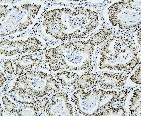
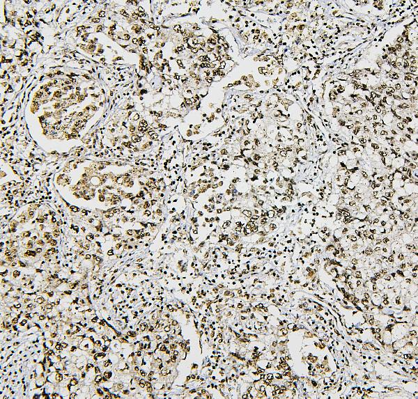
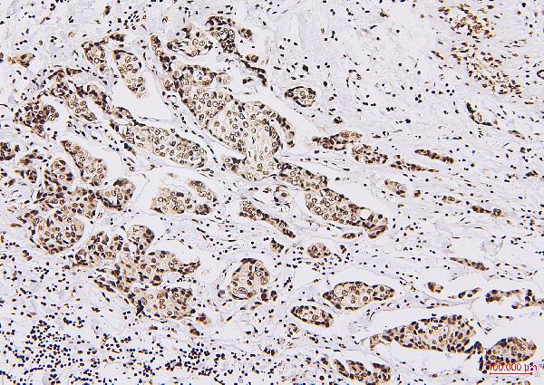
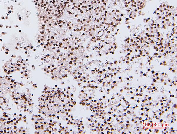
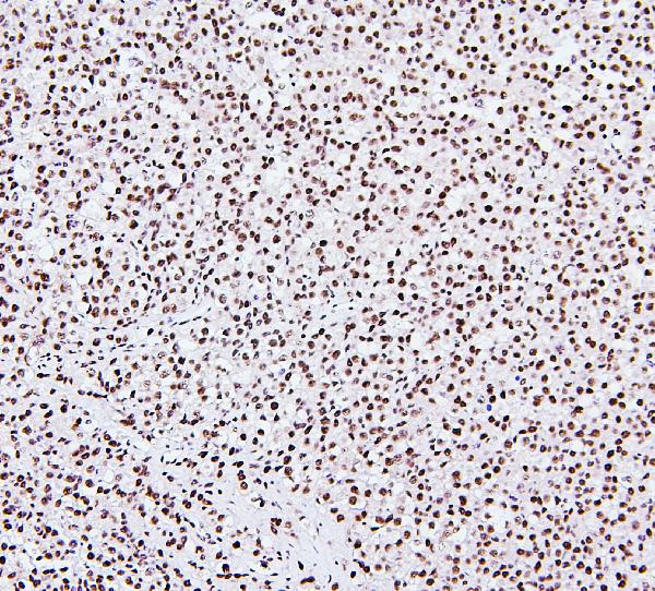
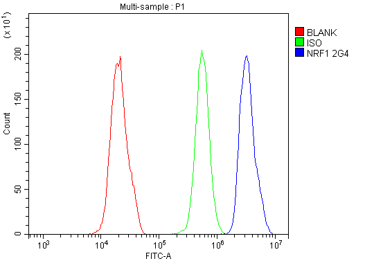
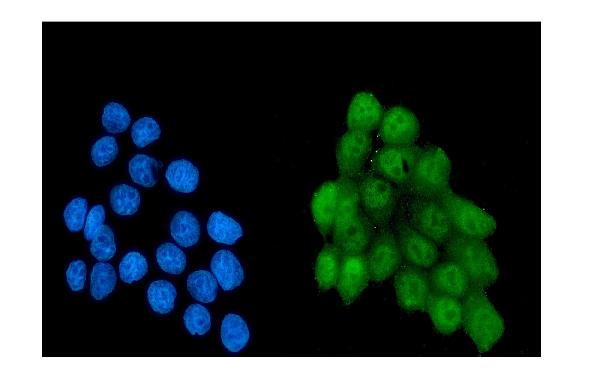
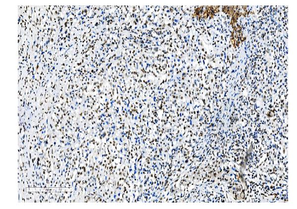
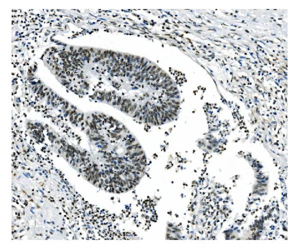
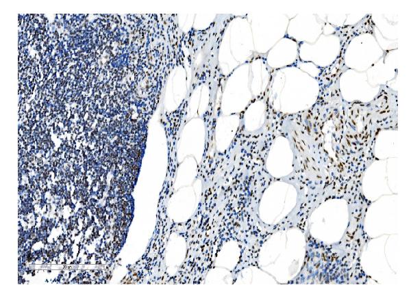
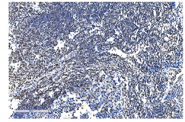
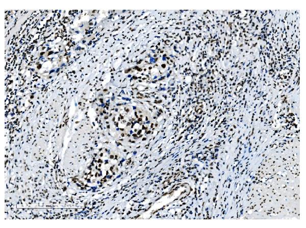
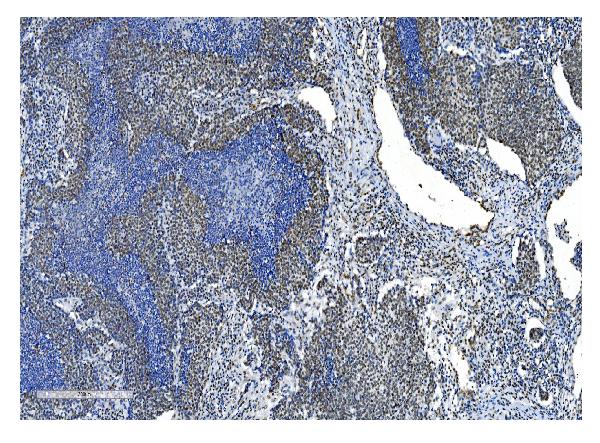
| Description | Anti-NRF1 Antibody Picoband™ (monoclonal, 2G4) . Tested in Flow Cytometry, IF, IHC, ICC, WB applications. This antibody reacts with Human. |
| Reconstitution | Add 0.2ml of distilled water will yield a concentration of 500 µg/ml. |
| Gene ID | 4899 |
|---|---|
| Other Names | Nuclear respiratory factor 1, NRF-1, Alpha palindromic-binding protein, Alpha-pal, NRF1 |
| Calculated MW | 54-70 kDa |
| Application Details | Western blot, 0.1-0.5 µg/ml, Human Immunohistochemistry (Paraffin-embedded Section), 0.5-1 µg/ml, Human, By Heat Immunocytochemistry/Immunofluorescence, 2 µg/ml, Human Flow Cytometry, 1-3 µg/1x10^6 cells, Human |
| Subcellular Localization | Nucleus. |
| Tissue Specificity | Ubiquitously expressed with strongest expression in skeletal muscle. |
| Contents | Each vial contains 4mg Trehalose, 0.9mg NaCl, 0.2mg Na2HPO4, 0.05mg NaN3. |
| Clone Names | Clone: 2G4 |
| Immunogen | E. coli-derived human NRF1 recombinant protein (Position: D246-Q503). |
| Cross Reactivity | No cross-reactivity with other proteins. |
| Storage | Store at -20˚C for one year from date of receipt. After reconstitution, at 4˚C for one month. It can also be aliquotted and stored frozen at -20˚C for six months. Avoid repeated freeze-thaw cycles. |
| Name | NRF1 |
|---|---|
| Function | Transcription factor that activates the expression of the EIF2S1 (EIF2-alpha) gene. Links the transcriptional modulation of key metabolic genes to cellular growth and development. Implicated in the control of nuclear genes required for respiration, heme biosynthesis, and mitochondrial DNA transcription and replication. |
| Cellular Location | Nucleus. |
| Tissue Location | Ubiquitously expressed with strongest expression in skeletal muscle |

Thousands of laboratories across the world have published research that depended on the performance of antibodies from Abcepta to advance their research. Check out links to articles that cite our products in major peer-reviewed journals, organized by research category.
info@abcepta.com, and receive a free "I Love Antibodies" mug.

Provided below are standard protocols that you may find useful for product applications.
Background
Nuclear respiratory factor 1, is also known as NRF1. This gene encodes a protein that homodimerizes and functions as a transcription factor which activates the expression of some key metabolic genes regulating cellular growth and nuclear genes required for respiration, heme biosynthesis, and mitochondrial DNA transcription and replication. The protein has also been associated with the regulation of neurite outgrowth. Alternative splicing results in multiple transcript variants. Confusion has occurred in bibliographic databases due to the shared symbol of NRF1 for this gene and for "nuclear factor (erythroid-derived 2)-like 1" which has an official symbol of NFE2L1.
If you have used an Abcepta product and would like to share how it has performed, please click on the "Submit Review" button and provide the requested information. Our staff will examine and post your review and contact you if needed.
If you have any additional inquiries please email technical services at tech@abcepta.com.
Ordering Information
Other Products
Shipping Information
