


 Foundational characteristics of cancer include proliferation, angiogenesis, migration, evasion of apoptosis, and cellular immortality. Find key markers for these cellular processes and antibodies to detect them.
 The SUMOplot™ Analysis Program predicts and scores sumoylation sites in your protein. SUMOylation is a post-translational modification involved in various cellular processes, such as nuclear-cytosolic transport, transcriptional regulation, apoptosis, protein stability, response to stress, and progression through the cell cycle.
 The Autophagy Receptor Motif Plotter predicts and scores autophagy receptor binding sites in your protein. Identifying proteins connected to this pathway is critical to understanding the role of autophagy in physiological as well as pathological processes such as development, differentiation, neurodegenerative diseases, stress, infection, and cancer.
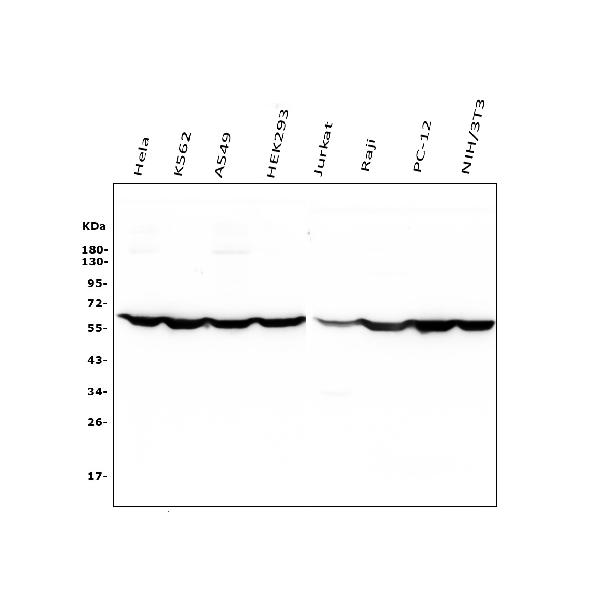
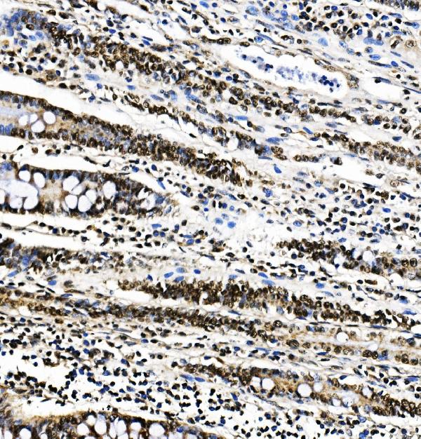
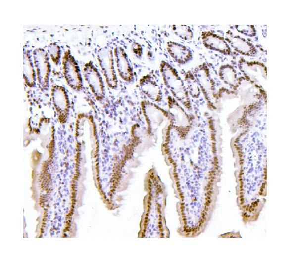
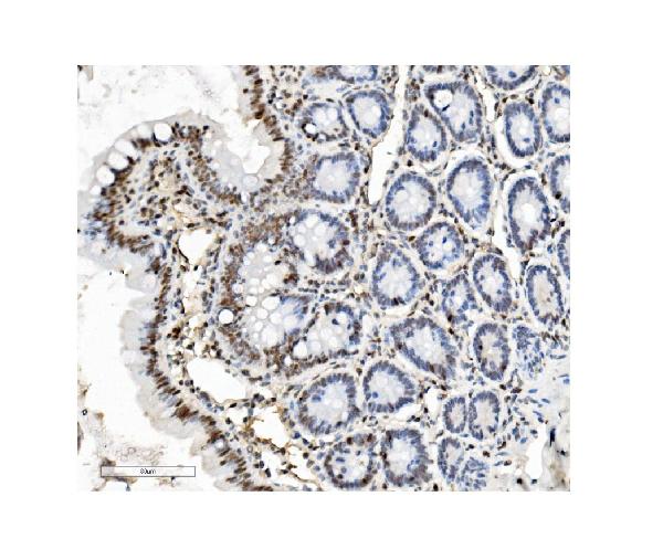
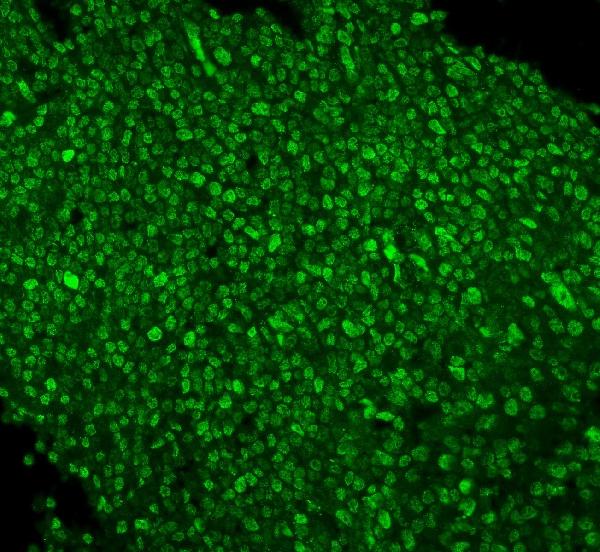
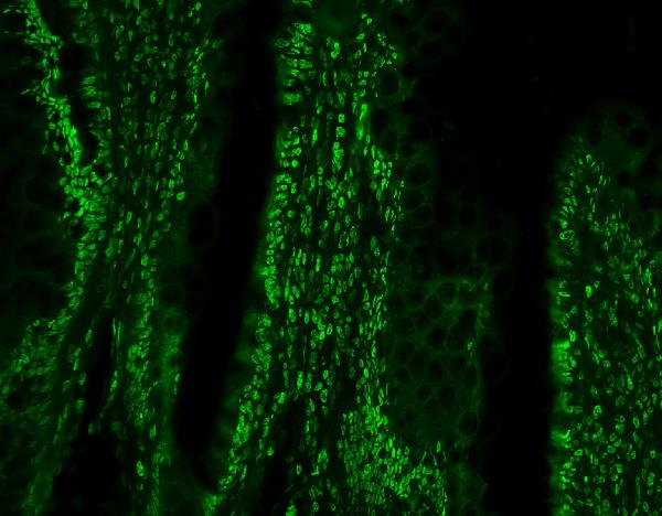
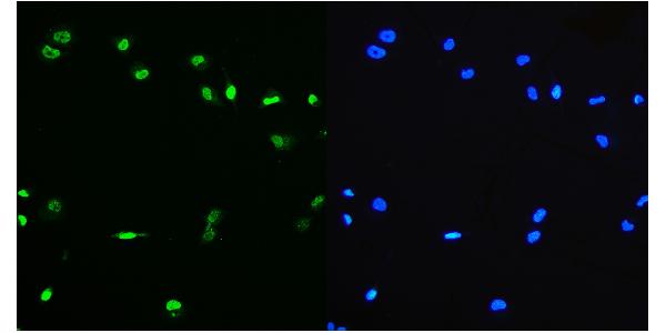
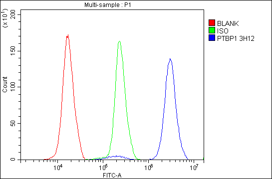
Anti-PTBP1 Antibody Picoband™(monoclonal, 3H12)
- SPECIFICATION
- CITATIONS
- PROTOCOLS
- BACKGROUND

Application
| WB, IHC, IF, ICC, FC |
|---|---|
| Primary Accession | P26599 |
| Host | Mouse |
| Isotype | Mouse IgG2b |
| Reactivity | Rat, Human, Mouse |
| Clonality | Monoclonal |
| Format | Lyophilized |
| Description | Anti-PTBP1 Antibody Picoband™ (monoclonal, 3H12) . Tested in Flow Cytometry, IF, IHC, ICC, WB applications. This antibody reacts with Human, Mouse, Rat. |
| Reconstitution | Add 0.2ml of distilled water will yield a concentration of 500 µg/ml. |
| Gene ID | 5725 |
|---|---|
| Other Names | Polypyrimidine tract-binding protein 1, PTB, 57 kDa RNA-binding protein PPTB-1, Heterogeneous nuclear ribonucleoprotein I, hnRNP I, PTBP1, PTB |
| Calculated MW | 57 kDa |
| Application Details | Western blot, 0.1-0.5 µg/ml, Human, Mouse, Rat Immunohistochemistry (Paraffin-embedded Section), 0.5-1 µg/ml, Human, Mouse, Rat Immunocytochemistry/Immunofluorescence, 2 µg/ml, Human Immunofluorescence, 2 µg/ml, Human, Rat Flow Cytometry, 1-3 µg/1x10^6 cells, Human |
| Protein Name | polypyrimidine tract binding protein 1 |
| Contents | Each vial contains 4mg Trehalose, 0.9mg NaCl, 0.2mg Na2HPO4, 0.05mg NaN3. |
| Clone Names | Clone: 3H12 |
| Immunogen | E.coli-derived human PTBP1 recombinant protein (Position: M1-A504). |
| Purification | Immunogen affinity purified. |
| Cross Reactivity | No cross-reactivity with other proteins. |
| Storage | Store at -20˚C for one year from date of receipt. After reconstitution, at 4˚C for one month. It can also be aliquotted and stored frozen at -20˚C for six months. Avoid repeated freeze-thaw cycles. |
| Name | PTBP1 |
|---|---|
| Synonyms | PTB |
| Function | Plays a role in pre-mRNA splicing and in the regulation of alternative splicing events. Activates exon skipping of its own pre- mRNA during muscle cell differentiation. Binds to the polypyrimidine tract of introns. May promote RNA looping when bound to two separate polypyrimidine tracts in the same pre-mRNA. May promote the binding of U2 snRNP to pre-mRNA. Cooperates with RAVER1 to modulate switching between mutually exclusive exons during maturation of the TPM1 pre- mRNA. Represses the splicing of MAPT/Tau exon 10 (PubMed:15009664). Binds to polypyrimidine-rich controlling element (PCE) of CFTR and promotes exon skipping of CFTR exon 9, thereby antagonizing TIA1 and its role in exon inclusion of CFTR exon 9 (PubMed:14966131). Plays a role in the splicing of pyruvate kinase PKM by binding repressively to a polypyrimidine tract flanking PKM exon 9, inhibiting exon 9 inclusion and resulting in exon 10 inclusion and production of the PKM M2 isoform (PubMed:20010808). In case of infection by picornaviruses, binds to the viral internal ribosome entry site (IRES) and stimulates the IRES- mediated translation (PubMed:21518806). |
| Cellular Location | Nucleus. |

Thousands of laboratories across the world have published research that depended on the performance of antibodies from Abcepta to advance their research. Check out links to articles that cite our products in major peer-reviewed journals, organized by research category.
info@abcepta.com, and receive a free "I Love Antibodies" mug.

Provided below are standard protocols that you may find useful for product applications.
Background
Polypyrimidine tract-binding protein 1 is a protein that in humans is encoded by the PTBP1 gene. It is mapped to 19p13.3. This gene belongs to the subfamily of ubiquitously expressed heterogeneous nuclear ribonucleoproteins (hnRNPs). The hnRNPs are RNA-binding proteins and they complex with heterogeneous nuclear RNA (hnRNA). These proteins are associated with pre-mRNAs in the nucleus and appear to influence pre-mRNA processing and other aspects of mRNA metabolism and transport. While all of the hnRNPs are present in the nucleus, some seem to shuttle between the nucleus and the cytoplasm. The hnRNP proteins have distinct nucleic acid binding properties. The protein encoded by this gene has four repeats of quasi-RNA recognition motif (RRM) domains that bind RNAs. This protein binds to the intronic polypyrimidine tracts that requires pre-mRNA splicing and acts via the protein degradation ubiquitin-proteasome pathway. It may also promote the binding of U2 snRNP to pre-mRNAs. This protein is localized in the nucleoplasm and it is also detected in the perinucleolar structure. Alternatively spliced transcript variants encoding different isoforms have been described.
If you have used an Abcepta product and would like to share how it has performed, please click on the "Submit Review" button and provide the requested information. Our staff will examine and post your review and contact you if needed.
If you have any additional inquiries please email technical services at tech@abcepta.com.
Ordering Information
Other Products
Shipping Information
