


 Foundational characteristics of cancer include proliferation, angiogenesis, migration, evasion of apoptosis, and cellular immortality. Find key markers for these cellular processes and antibodies to detect them.
 The SUMOplot™ Analysis Program predicts and scores sumoylation sites in your protein. SUMOylation is a post-translational modification involved in various cellular processes, such as nuclear-cytosolic transport, transcriptional regulation, apoptosis, protein stability, response to stress, and progression through the cell cycle.
 The Autophagy Receptor Motif Plotter predicts and scores autophagy receptor binding sites in your protein. Identifying proteins connected to this pathway is critical to understanding the role of autophagy in physiological as well as pathological processes such as development, differentiation, neurodegenerative diseases, stress, infection, and cancer.
Anti-CD79b Antibody Picoband™(monoclonal, 6H11)
- SPECIFICATION
- CITATIONS
- PROTOCOLS
- BACKGROUND

Application
| WB, IHC |
|---|---|
| Primary Accession | P40259 |
| Host | Mouse |
| Isotype | Mouse IgG1 |
| Reactivity | Rat, Human |
| Clonality | Monoclonal |
| Format | Lyophilized |
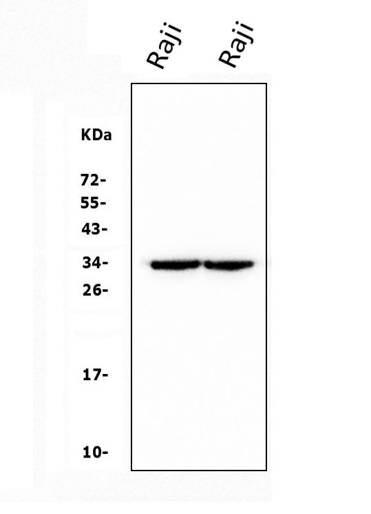
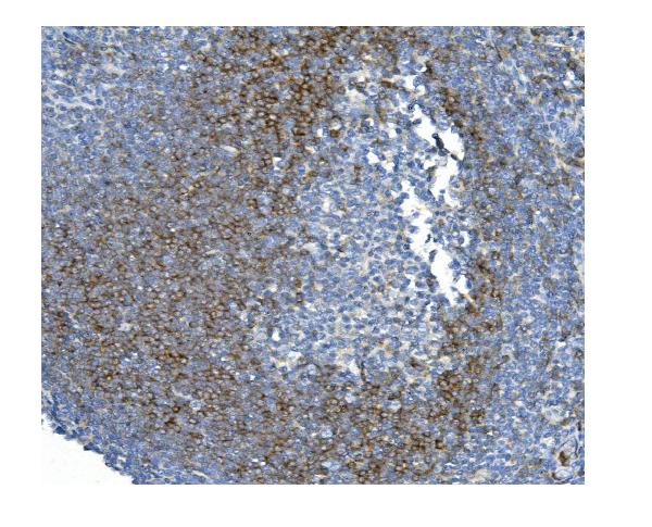
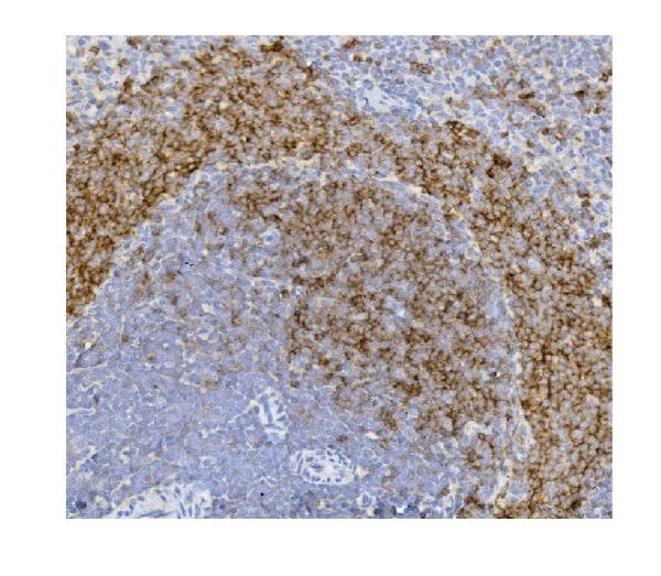
| Description | Anti-CD79b Antibody Picoband™ (monoclonal, 6H11) . Tested in IHC, WB applications. This antibody reacts with Human, Rat. |
| Reconstitution | Add 0.2ml of distilled water will yield a concentration of 500 µg/ml. |
| Gene ID | 974 |
|---|---|
| Other Names | B-cell antigen receptor complex-associated protein beta chain, B-cell-specific glycoprotein B29, Ig-beta, Immunoglobulin-associated B29 protein, CD79b, CD79B, B29, IGB |
| Calculated MW | 35 kDa |
| Application Details | Western blot, 0.1-0.5 µg/ml, Human Immunohistochemistry (Paraffin-embedded Section), 0.5-1 µg/ml, Human, Rat |
| Protein Name | CD79b molecule |
| Contents | Each vial contains 4mg Trehalose, 0.9mg NaCl, 0.2mg Na2HPO4, 0.05mg NaN3. |
| Clone Names | Clone: 6H11 |
| Immunogen | E.coli-derived human CD79b recombinant protein (Position: A29-E229). Human CD79b shares 70% amino acid (aa) sequence identity with mouse CD79b. |
| Purification | Immunogen affinity purified. |
| Cross Reactivity | No cross-reactivity with other proteins. |
| Storage | Store at -20˚C for one year from date of receipt. After reconstitution, at 4˚C for one month. It can also be aliquotted and stored frozen at -20˚C for six months. Avoid repeated freeze-thaw cycles. |
| Name | CD79B |
|---|---|
| Synonyms | B29, IGB |
| Function | Required in cooperation with CD79A for initiation of the signal transduction cascade activated by the B-cell antigen receptor complex (BCR) which leads to internalization of the complex, trafficking to late endosomes and antigen presentation. Enhances phosphorylation of CD79A, possibly by recruiting kinases which phosphorylate CD79A or by recruiting proteins which bind to CD79A and protect it from dephosphorylation. |
| Cellular Location | Cell membrane; Single-pass type I membrane protein. Note=Following antigen binding, the BCR has been shown to translocate from detergent-soluble regions of the cell membrane to lipid rafts although signal transduction through the complex can also occur outside lipid rafts. |
| Tissue Location | B-cells. |

Thousands of laboratories across the world have published research that depended on the performance of antibodies from Abcepta to advance their research. Check out links to articles that cite our products in major peer-reviewed journals, organized by research category.
info@abcepta.com, and receive a free "I Love Antibodies" mug.

Provided below are standard protocols that you may find useful for product applications.
Background
CD79b molecule, immunoglobulin-associated beta, also known as CD79B (Cluster of Differentiation 79B), is a human gene. By fluorescence in situ hybridization, It is mapped to 17q23.3. The CD79B protein together with the related CD79A protein, forms a dimer associated with membrane bound immunoglobulin in B-cells, thus forming the B-cell antigen receptor (BCR) which is a multimeric complex that includes the antigen-specific component, surface immunoglobulin (Ig). CD79b also can enhances phosphorylation of CD79A, possibly by recruiting kinases which phosphorylate CD79A or by recruiting proteins which bind to CD79A and protect it from dephosphorylation.
If you have used an Abcepta product and would like to share how it has performed, please click on the "Submit Review" button and provide the requested information. Our staff will examine and post your review and contact you if needed.
If you have any additional inquiries please email technical services at tech@abcepta.com.
Ordering Information
Other Products
Shipping Information
