


 Foundational characteristics of cancer include proliferation, angiogenesis, migration, evasion of apoptosis, and cellular immortality. Find key markers for these cellular processes and antibodies to detect them.
 The SUMOplot™ Analysis Program predicts and scores sumoylation sites in your protein. SUMOylation is a post-translational modification involved in various cellular processes, such as nuclear-cytosolic transport, transcriptional regulation, apoptosis, protein stability, response to stress, and progression through the cell cycle.
 The Autophagy Receptor Motif Plotter predicts and scores autophagy receptor binding sites in your protein. Identifying proteins connected to this pathway is critical to understanding the role of autophagy in physiological as well as pathological processes such as development, differentiation, neurodegenerative diseases, stress, infection, and cancer.
Anti-ch TOG/CKAP5 Antibody Picoband™ (monoclonal, 3C13)
- SPECIFICATION
- CITATIONS
- PROTOCOLS
- BACKGROUND

Application
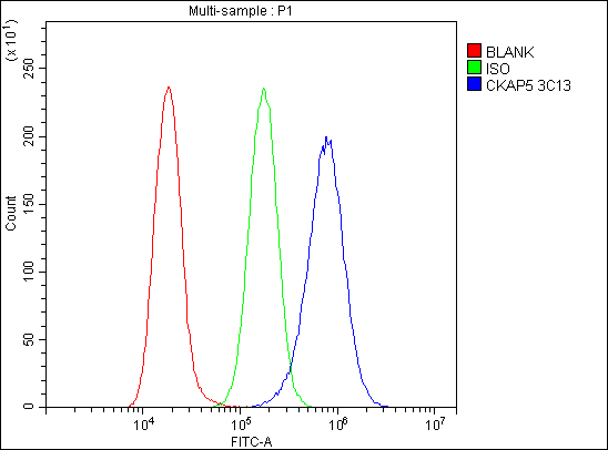
| WB, IHC, IF, ICC, FC |
|---|---|
| Primary Accession | Q14008 |
| Host | Mouse |
| Isotype | Mouse IgG2b |
| Reactivity | Rat, Human, Mouse |
| Clonality | Monoclonal |
| Format | Lyophilized |
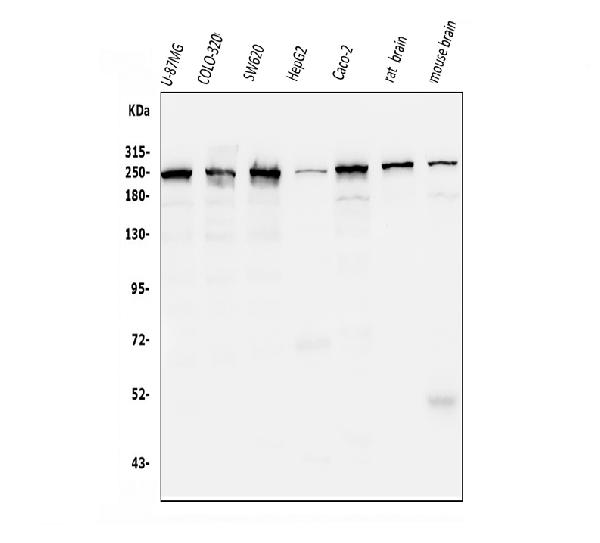
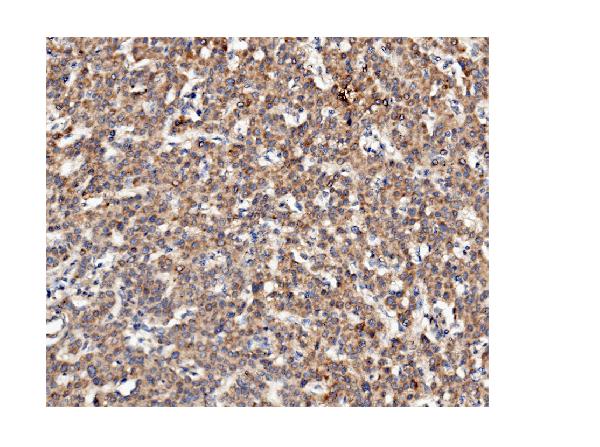
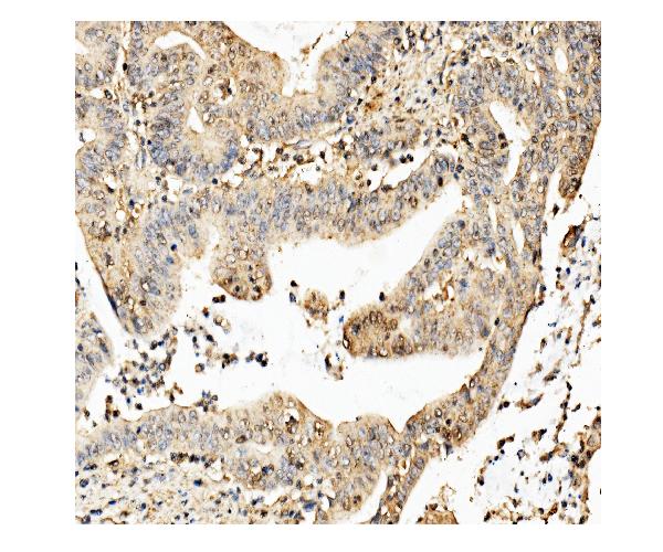
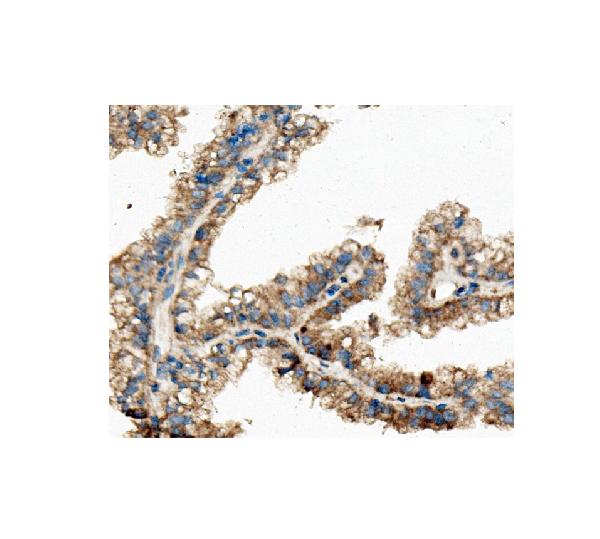
| Description | Anti-ch TOG/CKAP5 Antibody Picoband™ (monoclonal, 3C13) . Tested in Flow Cytometry, IF, IHC, ICC, WB applications. This antibody reacts with Human, Mouse, Rat. |
| Reconstitution | Add 0.2ml of distilled water will yield a concentration of 500ug/ml. |
| Gene ID | 9793 |
|---|---|
| Other Names | Cytoskeleton-associated protein 5, Colonic and hepatic tumor overexpressed gene protein, Ch-TOG, CKAP5, KIAA0097 |
| Calculated MW | 225 kDa |
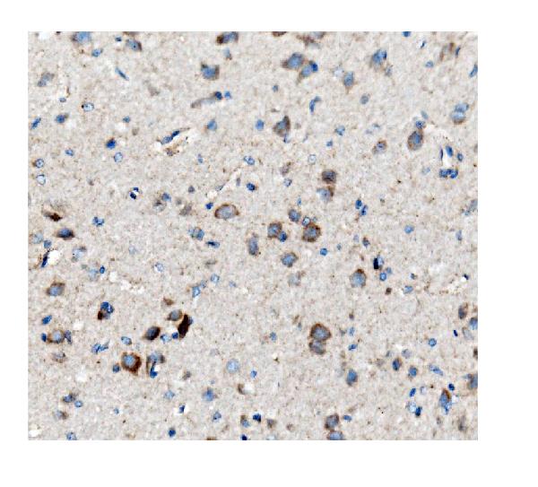
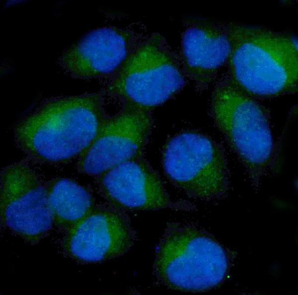
| Application Details | Western blot, 0.25-0.5 µg/ml, Human, Mouse, Rat Immunohistochemistry (Paraffin-embedded Section), 2-5 µg/ml, Human, Rat Immunocytochemistry/Immunofluorescence, 5 µg/ml, Human Flow Cytometry, 1-3 µg/1x10^6 cells, Human |
| Contents | Each vial contains 4mg Trehalose, 0.9mg NaCl and 0.2mg Na2HPO4. |
| Clone Names | Clone: 3C13 |
| Immunogen | E.coli-derived human ch TOG/CKAP5 recombinant protein (Position: M1-L221). |
| Purification | Immunogen affinity purified. |
| Storage | Store at -20˚C for one year from date of receipt. After reconstitution, at 4˚C for one month. It can also be aliquotted and stored frozen at -20˚C for six months. Avoid repeated freeze-thaw cycles. |
| Name | CKAP5 |
|---|---|
| Synonyms | KIAA0097 |
| Function | Binds to the plus end of microtubules and regulates microtubule dynamics and microtubule organization. Acts as a processive microtubule polymerase. Promotes cytoplasmic microtubule nucleation and elongation. Plays a major role in organizing spindle poles. In spindle formation protects kinetochore microtubules from depolymerization by KIF2C and has an essential role in centrosomal microtubule assembly independently of KIF2C activity. Contributes to centrosome integrity. Acts as a component of the TACC3/ch-TOG/clathrin complex proposed to contribute to stabilization of kinetochore fibers of the mitotic spindle by acting as inter-microtubule bridge. The TACC3/ch- TOG/clathrin complex is required for the maintenance of kinetochore fiber tension (PubMed:23532825). Enhances the strength of NDC80 complex-mediated kinetochore-tip microtubule attachments (PubMed:27156448). |
| Cellular Location | Cytoplasm, cytoskeleton, microtubule organizing center, centrosome. Cytoplasm, cytoskeleton, spindle pole. Cytoplasm, cytoskeleton, spindle. Chromosome, centromere, kinetochore. Note=Detected on centrosomes and kinetochores during interphase and mitosis independently from TACC3 and clathrin. Located to spindle poles and microtubules during mitosis. In complex with TACC3 localized to microtubule plus-ends in mitosis and interphase. In complex with TACC3 and clathrin localized to inter- microtubule bridges in mitotic spindles. Accumulation sites at microtubule plus ends protruded approximately 100 nm from MAPRE1/EB1 sites in interphase cells. |
| Tissue Location | Overexpressed in hepatomas and colonic tumors. Also expressed in skeletal muscle, brain, heart, placenta, lung, liver, kidney and pancreas. Expression is elevated in the brain; highly expressed in the Purkinje cell bodies of the cerebellum |

Thousands of laboratories across the world have published research that depended on the performance of antibodies from Abcepta to advance their research. Check out links to articles that cite our products in major peer-reviewed journals, organized by research category.
info@abcepta.com, and receive a free "I Love Antibodies" mug.

Provided below are standard protocols that you may find useful for product applications.
Background
Cytoskeleton-associated protein 5 is a microtubule-associated protein that in humans is encoded by the CKAP5 gene. It is mapped to 11p11.2. This gene encodes a cytoskeleton-associated protein which belongs to the TOG/XMAP215 family. The N-terminal half of this protein contains a microtubule-binding domain and the C-terminal half contains a KXGS motif for binding tubulin dimers. This protein has two distinct roles in spindle formation; it protects kinetochore microtubules from depolymerization and plays an essential role in centrosomal microtubule assembly. This protein may be necessary for the proper interaction of microtubules with the cell cortex for directional cell movement. It also plays a role in translation of the myelin basic protein (MBP) mRNA by interact ernatively spliced transcript variants encoding different isoforms have been identified.
If you have used an Abcepta product and would like to share how it has performed, please click on the "Submit Review" button and provide the requested information. Our staff will examine and post your review and contact you if needed.
If you have any additional inquiries please email technical services at tech@abcepta.com.
Ordering Information
Other Products
Shipping Information
