


 Foundational characteristics of cancer include proliferation, angiogenesis, migration, evasion of apoptosis, and cellular immortality. Find key markers for these cellular processes and antibodies to detect them.
 The SUMOplot™ Analysis Program predicts and scores sumoylation sites in your protein. SUMOylation is a post-translational modification involved in various cellular processes, such as nuclear-cytosolic transport, transcriptional regulation, apoptosis, protein stability, response to stress, and progression through the cell cycle.
 The Autophagy Receptor Motif Plotter predicts and scores autophagy receptor binding sites in your protein. Identifying proteins connected to this pathway is critical to understanding the role of autophagy in physiological as well as pathological processes such as development, differentiation, neurodegenerative diseases, stress, infection, and cancer.
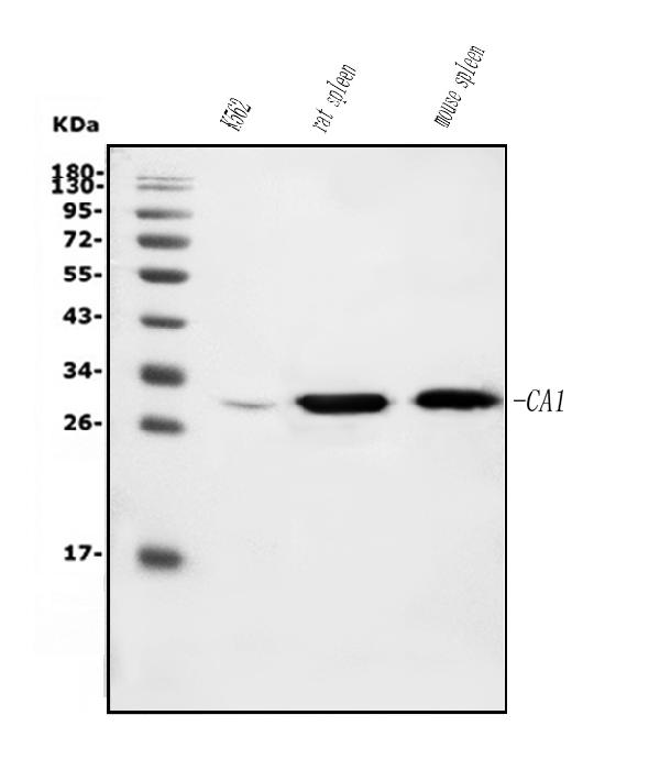
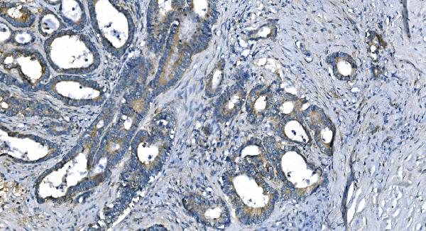
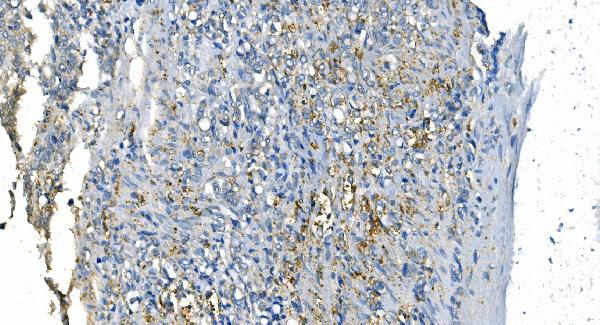
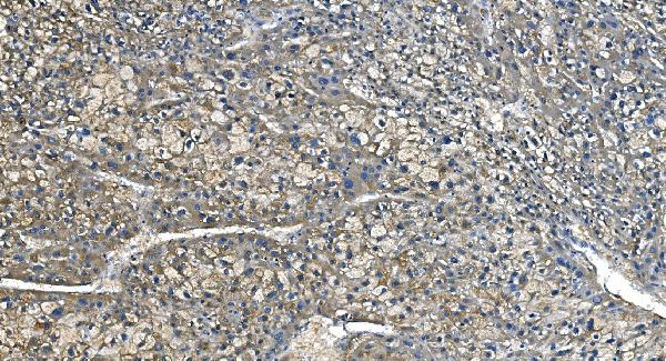
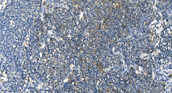
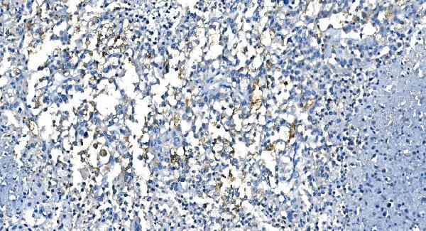
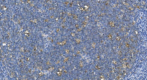
Anti-Carbonic Anhydrase I/CA1 Picoband™ Antibody (monoclonal, 2B5)
- SPECIFICATION
- CITATIONS
- PROTOCOLS
- BACKGROUND

Application
| WB, IHC |
|---|---|
| Primary Accession | P00915 |
| Host | Mouse |
| Isotype | Mouse IgG1 |
| Reactivity | Rat, Human, Mouse |
| Clonality | Monoclonal |
| Format | Lyophilized |
| Description | Anti-Carbonic Anhydrase I/CA1 Picoband™ Antibody (monoclonal, 2B5) . Tested in IHC, WB applications. This antibody reacts with Human, Mouse, Rat. |
| Reconstitution | Add 0.2ml of distilled water will yield a concentration of 500ug/ml. |
| Gene ID | 759 |
|---|---|
| Other Names | Carbonic anhydrase 1, 4.2.1.1, Carbonate dehydratase I, Carbonic anhydrase B, CAB, Carbonic anhydrase I, CA-I, Cyanamide hydratase CA1, 4.2.1.69, CA1 |
| Calculated MW | 29 kDa |
| Application Details | Western blot, 0.25-0.5 µg/ml, Human, Mouse, Rat Immunohistochemistry (Paraffin-embedded Section), 2-5 µg/ml, Human |
| Contents | Each vial contains 4mg Trehalose, 0.9mg NaCl and 0.2mg Na2HPO4. |
| Clone Names | Clone: 2B5 |
| Immunogen | E.coli-derived human CA1 recombinant protein (Position: D9-F261). Human CA1 shares 78.5% and 81% amino acid (aa) sequence identity with mouse and rat CA1, respectively. |
| Purification | Immunogen affinity purified. |
| Storage | Store at -20˚C for one year from date of receipt. After reconstitution, at 4˚C for one month. It can also be aliquotted and stored frozen at -20˚C for six months. Avoid repeated freeze-thaw cycles. |
| Name | CA1 |
|---|---|
| Function | Catalyzes the reversible hydration of carbon dioxide (PubMed:10550681, PubMed:16506782, PubMed:16686544, PubMed:16807956, PubMed:17127057, PubMed:17314045, PubMed:17407288, PubMed:18618712, PubMed:19186056, PubMed:19206230). Can hydrate cyanamide to urea (PubMed:10550681). |
| Cellular Location | Cytoplasm {ECO:0000250|UniProtKB:B0BNN3}. |

Thousands of laboratories across the world have published research that depended on the performance of antibodies from Abcepta to advance their research. Check out links to articles that cite our products in major peer-reviewed journals, organized by research category.
info@abcepta.com, and receive a free "I Love Antibodies" mug.

Provided below are standard protocols that you may find useful for product applications.
Background
Carbonic anhydrase 1 is an enzyme that in humans is encoded by the CA1 gene. It is a member of the Carbonic anhydrase. The CA1 gene is mapped to 8q22. CAI has got about 260 amino acids. This protein is highly expressed in erythrocytes. As catalysts of the reversible hydration of carbon dioxide, CAI participates in a variety of biologic processes like respiration, calcification, acid-base balance etc.
If you have used an Abcepta product and would like to share how it has performed, please click on the "Submit Review" button and provide the requested information. Our staff will examine and post your review and contact you if needed.
If you have any additional inquiries please email technical services at tech@abcepta.com.
Ordering Information
Other Products
Shipping Information
