


 Foundational characteristics of cancer include proliferation, angiogenesis, migration, evasion of apoptosis, and cellular immortality. Find key markers for these cellular processes and antibodies to detect them.
 The SUMOplot™ Analysis Program predicts and scores sumoylation sites in your protein. SUMOylation is a post-translational modification involved in various cellular processes, such as nuclear-cytosolic transport, transcriptional regulation, apoptosis, protein stability, response to stress, and progression through the cell cycle.
 The Autophagy Receptor Motif Plotter predicts and scores autophagy receptor binding sites in your protein. Identifying proteins connected to this pathway is critical to understanding the role of autophagy in physiological as well as pathological processes such as development, differentiation, neurodegenerative diseases, stress, infection, and cancer.
Anti-DDX1 Picoband™ Antibody (monoclonal, 2D4)

- SPECIFICATION
- CITATIONS
- PROTOCOLS
- BACKGROUND

Application
| WB, IHC, FC |
|---|---|
| Primary Accession | Q92499 |
| Host | Mouse |
| Isotype | Mouse IgG2a |
| Reactivity | Rat, Human, Mouse |
| Clonality | Monoclonal |
| Format | Lyophilized |
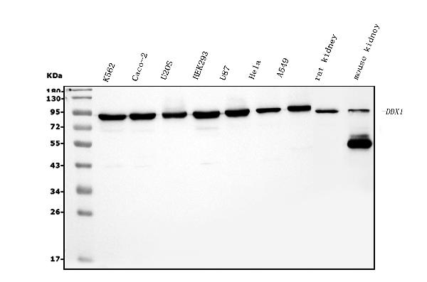
| Description | Anti-DDX1 Picoband™ Antibody (monoclonal, 2D4) . Tested in Flow Cytometry, IHC, WB applications. This antibody reacts with Human, Mouse, Rat. |
| Reconstitution | Add 0.2ml of distilled water will yield a concentration of 500ug/ml. |
| Gene ID | 1653 |
|---|---|
| Other Names | ATP-dependent RNA helicase DDX1, 3.6.4.13, DEAD box protein 1, DEAD box protein retinoblastoma, DBP-RB, DDX1 |
| Calculated MW | 86 kDa |
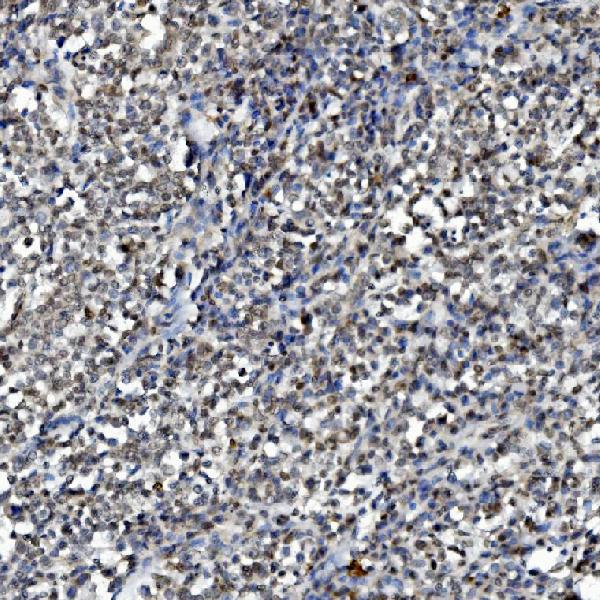
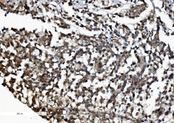
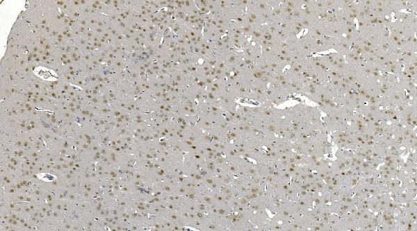
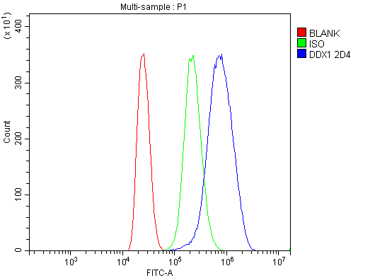
| Application Details | Western blot, 0.1-0.25 µg/ml, Human, Mouse, Rat Immunohistochemistry (Paraffin-embedded Section), 2-5 µg/ml, Human, Rat Flow Cytometry, 1-3 µg/1x10^6 cells, Human |
| Contents | Each vial contains 4mg Trehalose, 0.9mg NaCl and 0.2mg Na2HPO4. |
| Clone Names | Clone: 2D4 |
| Immunogen | E.coli-derived human DDX1 recombinant protein (Position: K562-F740). |
| Purification | Immunogen affinity purified. |
| Storage | Store at -20˚C for one year from date of receipt. After reconstitution, at 4˚C for one month. It can also be aliquotted and stored frozen at -20˚C for six months. Avoid repeated freeze-thaw cycles. |
| Name | DDX1 |
|---|---|
| Function | Acts as an ATP-dependent RNA helicase, able to unwind both RNA-RNA and RNA-DNA duplexes. Possesses 5' single-stranded RNA overhang nuclease activity. Possesses ATPase activity on various RNA, but not DNA polynucleotides. May play a role in RNA clearance at DNA double- strand breaks (DSBs), thereby facilitating the template-guided repair of transcriptionally active regions of the genome. Together with RELA, acts as a coactivator to enhance NF-kappa-B-mediated transcriptional activation. Acts as a positive transcriptional regulator of cyclin CCND2 expression. Binds to the cyclin CCND2 promoter region. Associates with chromatin at the NF-kappa-B promoter region via association with RELA. Binds to poly(A) RNA. May be involved in 3'-end cleavage and polyadenylation of pre-mRNAs. Component of the tRNA-splicing ligase complex required to facilitate the enzymatic turnover of catalytic subunit RTCB: together with archease (ZBTB8OS), acts by facilitating the guanylylation of RTCB, a key intermediate step in tRNA ligation (PubMed:24870230). Component of a multi-helicase-TICAM1 complex that acts as a cytoplasmic sensor of viral double-stranded RNA (dsRNA) and plays a role in the activation of a cascade of antiviral responses including the induction of pro-inflammatory cytokines via the adapter molecule TICAM1. Specifically binds (via helicase ATP-binding domain) on both short and long poly(I:C) dsRNA (By similarity). |
| Cellular Location | Nucleus. Cytoplasm. Cytoplasmic granule. Cytoplasm, cytosol {ECO:0000250|UniProtKB:Q91VR5}. Mitochondrion {ECO:0000250|UniProtKB:Q91VR5}. Note=Localized with MBNL1, TIAL1 and YBX1 in stress granules upon stress. Localized with CSTF2 in cleavage bodies. Forms large aggregates called DDX1 bodies. Relocalized into multiple foci (IR-induced foci or IRIF) after IR treatment, a process that depends on the presence of chromosomal DNA and/or RNA-DNA duplexes. Relocalized at sites of DNA double-strand breaks (DSBs) in an ATM-dependent manner after IR treatment. Colocalized with RELA in the nucleus upon TNF-alpha induction. Enters into the nucleus in case of active transcription while it accumulates in cytosol when transcription level is low (PubMed:24608264). Colocalizes in the cytosol with DDX21, DHX36 and TICAM1. Colocalizes in the mitochondria with TICAM1 and poly(I:C) RNA ligand. The multi-helicase-TICAM1 complex may translocate to the mitochondria upon poly(I:C) stimulation (By similarity) {ECO:0000250|UniProtKB:Q91VR5, ECO:0000269|PubMed:24608264} |
| Tissue Location | Highest levels of transcription in 2 retinoblastoma cell lines and in tissues of neuroectodermal origin including the retina, brain, and spinal cord. |

Thousands of laboratories across the world have published research that depended on the performance of antibodies from Abcepta to advance their research. Check out links to articles that cite our products in major peer-reviewed journals, organized by research category.
info@abcepta.com, and receive a free "I Love Antibodies" mug.

Provided below are standard protocols that you may find useful for product applications.
Background
ATP-dependent RNA helicase DDX1 is an enzyme that in humans is encoded by the DDX1 gene. It is mapped to 2p24.3. DEAD box proteins, characterized by the conserved motif Asp-Glu-Ala-Asp (DEAD), are putative RNA helicases. They are implicated in a number of cellular processes involving alteration of RNA secondary structure such as translation initiation, nuclear and mitochondrial splicing, and ribosome and spliceosome assembly. Based on their distribution patterns, some members of this family are believed to be involved in embryogenesis, spermatogenesis, and cellular growth and division. This gene encodes a DEAD box protein of unknown function. It shows high transcription levels in 2 retinoblastoma cell lines and in tissues of neuroectodermal origin.
If you have used an Abcepta product and would like to share how it has performed, please click on the "Submit Review" button and provide the requested information. Our staff will examine and post your review and contact you if needed.
If you have any additional inquiries please email technical services at tech@abcepta.com.
Ordering Information
Other Products
Shipping Information
