


 Foundational characteristics of cancer include proliferation, angiogenesis, migration, evasion of apoptosis, and cellular immortality. Find key markers for these cellular processes and antibodies to detect them.
 The SUMOplot™ Analysis Program predicts and scores sumoylation sites in your protein. SUMOylation is a post-translational modification involved in various cellular processes, such as nuclear-cytosolic transport, transcriptional regulation, apoptosis, protein stability, response to stress, and progression through the cell cycle.
 The Autophagy Receptor Motif Plotter predicts and scores autophagy receptor binding sites in your protein. Identifying proteins connected to this pathway is critical to understanding the role of autophagy in physiological as well as pathological processes such as development, differentiation, neurodegenerative diseases, stress, infection, and cancer.
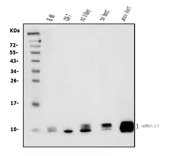
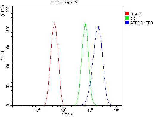
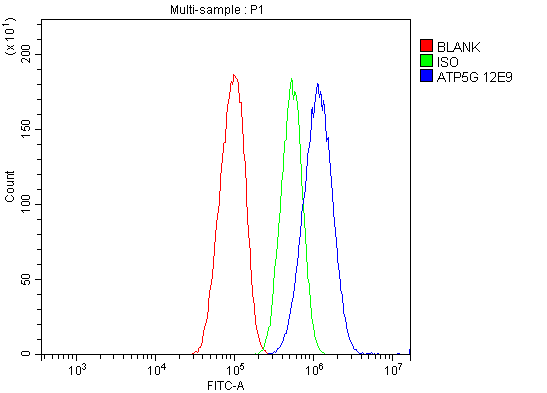
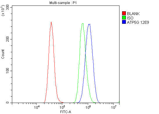
Anti-ATP5F1,2,3/ATP5MC1,2,3 Picoband™ Antibody (monoclonal, 12E9)
- SPECIFICATION
- CITATIONS
- PROTOCOLS
- BACKGROUND

Application
| WB, FC |
|---|---|
| Primary Accession | P05496 |
| Host | Mouse |
| Isotype | Mouse IgG2b |
| Reactivity | Rat, Human, Mouse, Monkey |
| Clonality | Monoclonal |
| Format | Lyophilized |
| Description | Anti-ATP5F1,2,3/ATP5MC1,2,3 Picoband™ Antibody (monoclonal, 12E9) . Tested in Flow Cytometry, WB applications. This antibody reacts with Human, Monkey, Mouse, Rat. |
| Reconstitution | Add 0.2ml of distilled water will yield a concentration of 500ug/ml. |
| Gene ID | 516 |
|---|---|
| Other Names | ATP synthase F(0) complex subunit C1, mitochondrial, ATP synthase lipid-binding protein, ATP synthase membrane subunit c locus 1 {ECO:0000312|HGNC:HGNC:841}, ATP synthase proteolipid P1, ATP synthase proton-transporting mitochondrial F(0) complex subunit C1, ATPase protein 9, ATPase subunit c, ATP5MC1 (HGNC:841) |
| Calculated MW | 10-14 kDa |
| Application Details | Western blot, 0.25-0.5 µg/ml, Human, Mouse, Rat, Monkey Flow Cytometry, 1-3 µg/1x10^6 cells, Human, Mouse, Rat |
| Contents | Each vial contains 4mg Trehalose, 0.9mg NaCl and 0.2mg Na2HPO4. |
| Clone Names | Clone: 12E9 |
| Immunogen | E.coli-derived human ATP5G1,2,3/ATP5MC1,2,3 recombinant protein (Position: D62-L113). |
| Purification | Immunogen affinity purified. |
| Storage | Store at -20˚C for one year from date of receipt. After reconstitution, at 4˚C for one month. It can also be aliquotted and stored frozen at -20˚C for six months. Avoid repeated freeze-thaw cycles. |
| Name | ATP5MC1 (HGNC:841) |
|---|---|
| Function | Subunit c, of the mitochondrial membrane ATP synthase complex (F(1)F(0) ATP synthase or Complex V) that produces ATP from ADP in the presence of a proton gradient across the membrane which is generated by electron transport complexes of the respiratory chain (Probable). ATP synthase complex consist of a soluble F(1) head domain - the catalytic core - and a membrane F(1) domain - the membrane proton channel (PubMed:37244256). These two domains are linked by a central stalk rotating inside the F(1) region and a stationary peripheral stalk (PubMed:37244256). During catalysis, ATP synthesis in the catalytic domain of F(1) is coupled via a rotary mechanism of the central stalk subunits to proton translocation (Probable). With the subunit a (MT- ATP6), forms the proton-conducting channel in the F(0) domain, that contains two crucial half-channels (inlet and outlet) that facilitate proton movement from the mitochondrial intermembrane space (IMS) into the matrix (PubMed:37244256). Protons are taken up via the inlet half- channel and released through the outlet half-channel, following a Grotthuss mechanism (PubMed:37244256). |
| Cellular Location | Mitochondrion membrane; Multi-pass membrane protein |

Thousands of laboratories across the world have published research that depended on the performance of antibodies from Abcepta to advance their research. Check out links to articles that cite our products in major peer-reviewed journals, organized by research category.
info@abcepta.com, and receive a free "I Love Antibodies" mug.

Provided below are standard protocols that you may find useful for product applications.
Background
The ATP5MC1 gene is one of three human paralogs that encode membrane subunit c of the mitochondrial ATP synthase. It is mapped to 17q21.32. This gene encodes a subunit of mitochondrial ATP synthase. Mitochondrial ATP synthase catalyzes ATP synthesis, utilizing an electrochemical gradient of protons across the inner membrane during oxidative phosphorylation. ATP synthase is composed of two linked multi-subunit complexes: the soluble catalytic core, F1, and the membrane-spanning component, Fo, comprising the proton channel. The catalytic portion of mitochondrial ATP synthase consists of 5 different subunits (alpha, beta, gamma, delta, and epsilon) assembled with a stoichiometry of 3 alpha, 3 beta, and a single representative of the other 3. The proton channel seems to have nine subunits (a, b, c, d, e, f, g, F6 and 8). This gene is one of three genes that encode subunit c of the proton channel. Each of the three genes have distinct mitochondrial import sequences but encode the identical mature protein. Alternatively spliced transcript variants encoding the same protein have been identified.
If you have used an Abcepta product and would like to share how it has performed, please click on the "Submit Review" button and provide the requested information. Our staff will examine and post your review and contact you if needed.
If you have any additional inquiries please email technical services at tech@abcepta.com.
Ordering Information
Other Products
Shipping Information
