


 Foundational characteristics of cancer include proliferation, angiogenesis, migration, evasion of apoptosis, and cellular immortality. Find key markers for these cellular processes and antibodies to detect them.
 The SUMOplot™ Analysis Program predicts and scores sumoylation sites in your protein. SUMOylation is a post-translational modification involved in various cellular processes, such as nuclear-cytosolic transport, transcriptional regulation, apoptosis, protein stability, response to stress, and progression through the cell cycle.
 The Autophagy Receptor Motif Plotter predicts and scores autophagy receptor binding sites in your protein. Identifying proteins connected to this pathway is critical to understanding the role of autophagy in physiological as well as pathological processes such as development, differentiation, neurodegenerative diseases, stress, infection, and cancer.
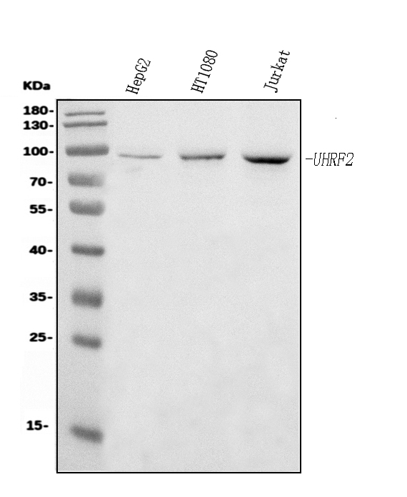
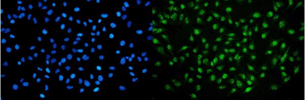
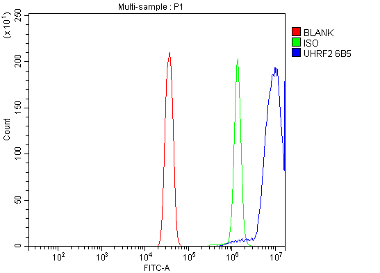
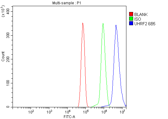
Anti-NIRF Antibody Picoband™ (monoclonal, 6B5)
- SPECIFICATION
- CITATIONS
- PROTOCOLS
- BACKGROUND

Application
| WB, IF, ICC, FC |
|---|---|
| Primary Accession | Q96PU4 |
| Host | Mouse |
| Isotype | Mouse IgG2b |
| Reactivity | Rat, Human |
| Clonality | Monoclonal |
| Format | Lyophilized |
| Description | Anti-NIRF Antibody Picoband™ (monoclonal, 6B5) . Tested in Flow Cytometry, IF, ICC, WB applications. This antibody reacts with Human, Rat. |
| Reconstitution | Adding 0.2 ml of distilled water will yield a concentration of 500 µg/ml. |
| Gene ID | 115426 |
|---|---|
| Other Names | E3 ubiquitin-protein ligase UHRF2, 2.3.2.27, Np95/ICBP90-like RING finger protein, Np95-like RING finger protein, Nuclear protein 97, Nuclear zinc finger protein Np97, RING finger protein 107, RING-type E3 ubiquitin transferase UHRF2, Ubiquitin-like PHD and RING finger domain-containing protein 2, Ubiquitin-like-containing PHD and RING finger domains protein 2, UHRF2, NIRF, RNF107 |
| Calculated MW | 90 kDa |
| Application Details | Western blot, 0.25-0.5 µg/ml, Human Immunocytochemistry/Immunofluorescence, 5 µg/ml, Human Flow Cytometry, 1-3 µg/1x10^6 cells, Human, Rat |
| Contents | Each vial contains 4 mg Trehalose, 0.9 mg NaCl and 0.2 mg Na2HPO4. |
| Clone Names | Clone: 6B5 |
| Immunogen | A synthetic peptide corresponding to a sequence at the N-terminus of human NIRF, identical to the related mouse and rat sequences. |
| Purification | Immunogen affinity purified. |
| Storage | At -20°C for one year from date of receipt. After reconstitution, at 4°C for one month. It can also be aliquotted and stored frozen at -20°C for six months. Avoid repeated freezing and thawing. |
| Name | UHRF2 |
|---|---|
| Synonyms | NIRF, RNF107 |
| Function | E3 ubiquitin ligase that plays important roles in DNA methylation, histone modifications, cell cycle and DNA repair (PubMed:15178429, PubMed:23404503, PubMed:27743347, PubMed:29506131). Acts as a specific reader for 5-hydroxymethylcytosine (5hmC) and thereby recruits various substrates to these sites to ubiquitinate them (PubMed:24813944, PubMed:27129234). This activity also allows the maintenance of 5mC levels at specific genomic loci and regulates neuron-related gene expression (By similarity). Participates in cell cycle regulation by ubiquitinating cyclins CCND1 and CCNE1 and thereby inducing G1 arrest (PubMed:15178429, PubMed:15361834, PubMed:21952639). Also ubiquitinates PCNP leading to its degradation by the proteasome (PubMed:12176013, PubMed:14741369). Plays an active role in DNA damage repair by ubiquitinating p21/CDKN1A leading to its proteasomal degradation (PubMed:29923055). Also promotes DNA repair by acting as an interstrand cross-links (ICLs) sensor. Mechanistically, cooperates with UHRF1 to ensure recruitment of FANCD2 to ICLs, leading to FANCD2 monoubiquitination and subsequent activation (PubMed:30335751). Contributes to UV-induced DNA damage response by physically interacting with ATR in response to irradiation, thereby promoting ATR activation (PubMed:33848395). |
| Cellular Location | Nucleus {ECO:0000255|PROSITE-ProRule:PRU00358, ECO:0000269|PubMed:12176013, ECO:0000269|PubMed:23404503, ECO:0000269|PubMed:27129234, ECO:0000269|PubMed:27743347, ECO:0000269|PubMed:29923055, ECO:0000269|PubMed:30335751}. Chromosome. Note=Enriched at genomic loci that are enriched for 5-hydroxymethylcytosine (5hmC) |

Thousands of laboratories across the world have published research that depended on the performance of antibodies from Abcepta to advance their research. Check out links to articles that cite our products in major peer-reviewed journals, organized by research category.
info@abcepta.com, and receive a free "I Love Antibodies" mug.

Provided below are standard protocols that you may find useful for product applications.
Background
E3 ubiquitin-protein ligase UHRF2 is an enzyme that in humans is encoded by the UHRF2 gene. This gene encodes a nuclear protein which is involved in cell-cycle regulation. The encoded protein is a ubiquitin-ligase capable of ubiquinating PCNP (PEST-containing nuclear protein), and together they may play a role in tumorigenesis. The encoded protein contains an NIRF_N domain, a PHD finger, a set- and ring-associated (SRA) domain, and a RING finger domain and several of these domains have been shown to be essential for the regulation of cell proliferation. This protein may also have a role in intranuclear degradation of polyglutamine aggregates. Alternative splicing results in multiple transcript variants some of which are non-protein coding.
If you have used an Abcepta product and would like to share how it has performed, please click on the "Submit Review" button and provide the requested information. Our staff will examine and post your review and contact you if needed.
If you have any additional inquiries please email technical services at tech@abcepta.com.
Ordering Information
Other Products
Shipping Information
