


 Foundational characteristics of cancer include proliferation, angiogenesis, migration, evasion of apoptosis, and cellular immortality. Find key markers for these cellular processes and antibodies to detect them.
 The SUMOplot™ Analysis Program predicts and scores sumoylation sites in your protein. SUMOylation is a post-translational modification involved in various cellular processes, such as nuclear-cytosolic transport, transcriptional regulation, apoptosis, protein stability, response to stress, and progression through the cell cycle.
 The Autophagy Receptor Motif Plotter predicts and scores autophagy receptor binding sites in your protein. Identifying proteins connected to this pathway is critical to understanding the role of autophagy in physiological as well as pathological processes such as development, differentiation, neurodegenerative diseases, stress, infection, and cancer.
Anti-Moesin/MSN Antibody Picoband™ (monoclonal, 8D4)
- SPECIFICATION
- CITATIONS
- PROTOCOLS
- BACKGROUND

Application
| WB, IHC, IF, ICC, FC |
|---|---|
| Primary Accession | P26038 |
| Host | Mouse |
| Isotype | Mouse IgG1 |
| Reactivity | Rat, Human, Mouse, Monkey |
| Clonality | Monoclonal |
| Format | Lyophilized |
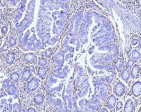
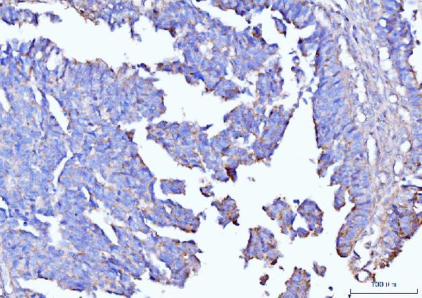
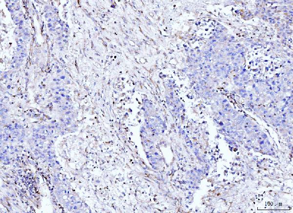
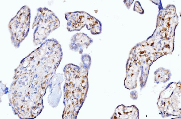
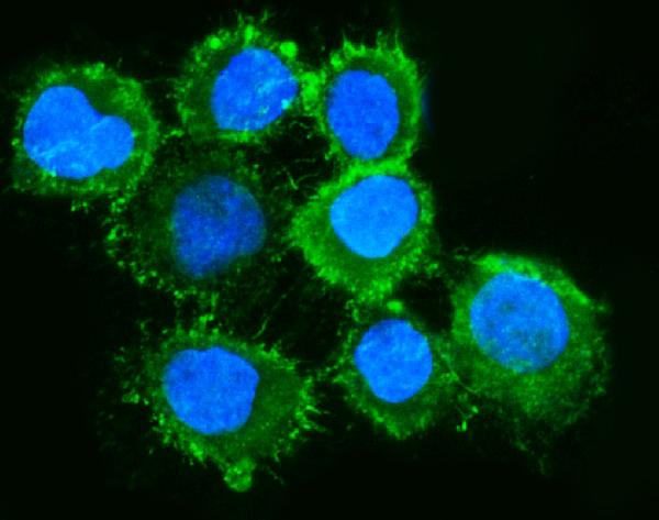
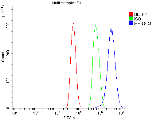
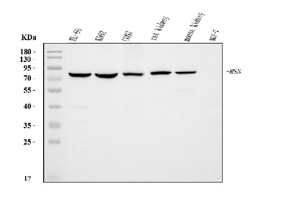
| Description | Anti-Moesin/MSN Antibody Picoband™ (monoclonal, 8D4) . Tested in FCM, IF, IHC, ICC, WB applications. This antibody reacts with Human, Mouse, Rat, Monkey. |
| Reconstitution | Adding 0.2 ml of distilled water will yield a concentration of 500 µg/ml. |
| Gene ID | 4478 |
|---|---|
| Other Names | Moesin, Membrane-organizing extension spike protein, MSN (HGNC:7373) |
| Calculated MW | 78 kDa |
| Application Details | Western blot, 0.25-0.5 µg/ml, Human, Mouse, Rat, Monkey Immunohistochemistry(Paraffin-embedded Section), 2-5 µg/ml, Human Immunocytochemistry/Immunofluorescence, 5 µg/ml, Human Flow Cytometry, 1-3 µg/1x^6 cells, Human |
| Contents | Each vial contains 4 mg Trehalose, 0.9 mg NaCl and 0.2 mg Na2HPO4. |
| Clone Names | Clone: 8D4 |
| Immunogen | E.coli-derived human Moesin/MSN recombinant protein (Position: R184-K568). |
| Purification | Immunogen affinity purified. |
| Storage | At -20°C for one year from date of receipt. After reconstitution, at 4°C for one month. It can also be aliquotted and stored frozen at -20°C for six months. Avoid repeated freezing and thawing. |
| Name | MSN (HGNC:7373) |
|---|---|
| Function | Ezrin-radixin-moesin (ERM) family protein that connects the actin cytoskeleton to the plasma membrane and thereby regulates the structure and function of specific domains of the cell cortex. Tethers actin filaments by oscillating between a resting and an activated state providing transient interactions between moesin and the actin cytoskeleton (PubMed:10212266). Once phosphorylated on its C-terminal threonine, moesin is activated leading to interaction with F-actin and cytoskeletal rearrangement (PubMed:10212266). These rearrangements regulate many cellular processes, including cell shape determination, membrane transport, and signal transduction (PubMed:12387735, PubMed:15039356). The role of moesin is particularly important in immunity acting on both T and B-cells homeostasis and self-tolerance, regulating lymphocyte egress from lymphoid organs (PubMed:9298994, PubMed:9616160). Modulates phagolysosomal biogenesis in macrophages (By similarity). Also participates in immunologic synapse formation (PubMed:27405666). |
| Cellular Location | Cell membrane; Peripheral membrane protein {ECO:0000250|UniProtKB:P26041}; Cytoplasmic side {ECO:0000250|UniProtKB:P26041}. Cytoplasm, cytoskeleton {ECO:0000250|UniProtKB:P26041}. Apical cell membrane {ECO:0000250|UniProtKB:P26041}; Peripheral membrane protein {ECO:0000250|UniProtKB:P26041}; Cytoplasmic side {ECO:0000250|UniProtKB:P26041}. Cell projection, microvillus membrane {ECO:0000250|UniProtKB:P26041}; Peripheral membrane protein {ECO:0000250|UniProtKB:P26041}; Cytoplasmic side {ECO:0000250|UniProtKB:P26041}. Cell projection, microvillus {ECO:0000250|UniProtKB:P26041}. Note=Phosphorylated form is enriched in microvilli-like structures at apical membrane. Increased cell membrane localization of both phosphorylated and non-phosphorylated forms seen after thrombin treatment (By similarity). Localizes at the uropods of T lymphoblasts. {ECO:0000250|UniProtKB:P26041, ECO:0000269|PubMed:18586956, ECO:0000269|PubMed:9298994} |
| Tissue Location | In all tissues and cultured cells studied. |

Thousands of laboratories across the world have published research that depended on the performance of antibodies from Abcepta to advance their research. Check out links to articles that cite our products in major peer-reviewed journals, organized by research category.
info@abcepta.com, and receive a free "I Love Antibodies" mug.

Provided below are standard protocols that you may find useful for product applications.
Background
Moesin is a protein that in humans is encoded by the MSN gene. It is mapped to Xq12. Moesin (for membrane-organizing extension spike protein) is a member of the ERM family which includes ezrin and radixin. ERM proteins appear to function as cross-linkers between plasma membranes and actin-based cytoskeletons. Moesin is localized to filopodia and other membranous protrusions that are important for cell-cell recognition and signaling and for cell movement.
If you have used an Abcepta product and would like to share how it has performed, please click on the "Submit Review" button and provide the requested information. Our staff will examine and post your review and contact you if needed.
If you have any additional inquiries please email technical services at tech@abcepta.com.
Ordering Information
Other Products
Shipping Information
