


 Foundational characteristics of cancer include proliferation, angiogenesis, migration, evasion of apoptosis, and cellular immortality. Find key markers for these cellular processes and antibodies to detect them.
 The SUMOplot™ Analysis Program predicts and scores sumoylation sites in your protein. SUMOylation is a post-translational modification involved in various cellular processes, such as nuclear-cytosolic transport, transcriptional regulation, apoptosis, protein stability, response to stress, and progression through the cell cycle.
 The Autophagy Receptor Motif Plotter predicts and scores autophagy receptor binding sites in your protein. Identifying proteins connected to this pathway is critical to understanding the role of autophagy in physiological as well as pathological processes such as development, differentiation, neurodegenerative diseases, stress, infection, and cancer.
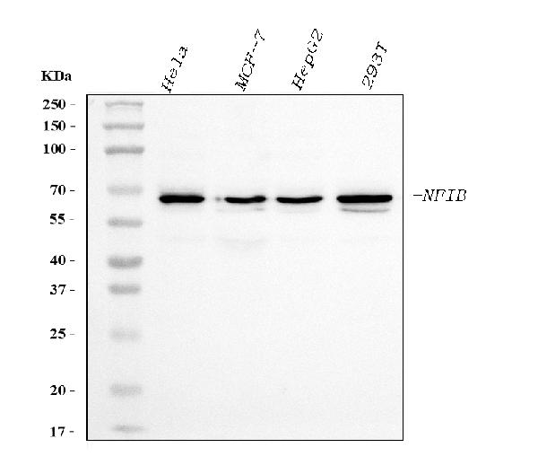
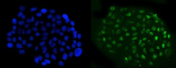
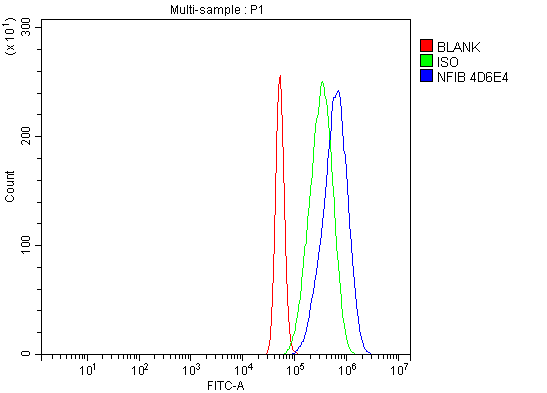
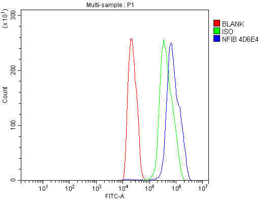
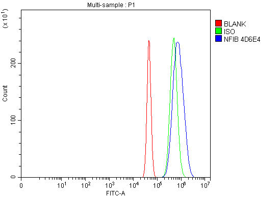
Anti-NFIB/NF1B2 Antibody Picoband™ (monoclonal, 4D6E4)
- SPECIFICATION
- CITATIONS
- PROTOCOLS
- BACKGROUND

Application
| WB, IF, ICC, FC |
|---|---|
| Primary Accession | O00712 |
| Host | Mouse |
| Isotype | Mouse IgG2b |
| Reactivity | Rat, Human, Mouse |
| Clonality | Monoclonal |
| Format | Lyophilized |
| Description | Anti-NFIB/NF1B2 Antibody Picoband™ (monoclonal, 4D6E4) . Tested in Flow Cytometry, IF, ICC, WB applications. This antibody reacts with Human, Mouse, Rat. |
| Reconstitution | Adding 0.2 ml of distilled water will yield a concentration of 500 µg/ml. |
| Gene ID | 4781 |
|---|---|
| Other Names | Nuclear factor 1 B-type, NF1-B, Nuclear factor 1/B, CCAAT-box-binding transcription factor, CTF, Nuclear factor I/B, NF-I/B, NFI-B, TGGCA-binding protein, NFIB |
| Calculated MW | 68 kDa |
| Application Details | Western blot, 0.25-0.5 µg/ml, Human Immunocytochemistry/Immunofluorescence, 5 µg/ml, Human Flow Cytometry, 1-3 µg/1x10^6 cells, Human, Mouse, Rat |
| Contents | Each vial contains 4 mg Trehalose, 0.9 mg NaCl and 0.2 mg Na2HPO4. |
| Clone Names | Clone: 4D6E4 |
| Immunogen | A synthetic peptide corresponding to a sequence in the middle region of human NFIB/NF1B2, identical to the related mouse and rat sequences. |
| Purification | Immunogen affinity purified. |
| Storage | At -20°C for one year from date of receipt. After reconstitution, at 4°C for one month. It can also be aliquotted and stored frozen at -20°C for six months. Avoid repeated freezing and thawing. |
| Name | NFIB |
|---|---|
| Function | Transcriptional activator of GFAP, essential for proper brain development (PubMed:30388402). Recognizes and binds the palindromic sequence 5'-TTGGCNNNNNGCCAA-3' present in viral and cellular promoters and in the origin of replication of adenovirus type 2. These proteins are individually capable of activating transcription and replication. |
| Cellular Location | Nucleus. |

Thousands of laboratories across the world have published research that depended on the performance of antibodies from Abcepta to advance their research. Check out links to articles that cite our products in major peer-reviewed journals, organized by research category.
info@abcepta.com, and receive a free "I Love Antibodies" mug.

Provided below are standard protocols that you may find useful for product applications.
Background
Nuclear factor 1 B-type is a protein that in humans is encoded by the NFIB gene. The NFIB gene is a part of the NFI gene complex that includes three other genes (NFIA, NFIC and NFIX). The NFIB gene is a protein coding gene that also serves as a transcription factor. This gene is essential in embryonic development and it works together with its gene complex to initiate tissue differentiation in the fetus. Through knockout experiments, researchers found that mice without the NFIB gene have severely underdeveloped lungs. This mutation does not seem to cause spontaneous abortions because in utero the fetus does not use its lungs for respiration. However, this becomes lethal once the fetus is born and has to take its first breath. It is thought that NFIB plays a role in down regulating the transcription factors TGF-β1 and Shh in normal gestation because they remained high in knockout experiments. The absence of NFIB also leads to insufficient amounts of surfactant being produced which is one reason why the mice cannot breathe once it is born. The knockout experiments demonstrated that NFIB has a significant role in fore-brain development. NFIB is typically found in pontine nuclei of the CNS, the cerebral cortex and the white matter of the brain and without NFIB these areas are dramatically affected.
If you have used an Abcepta product and would like to share how it has performed, please click on the "Submit Review" button and provide the requested information. Our staff will examine and post your review and contact you if needed.
If you have any additional inquiries please email technical services at tech@abcepta.com.
Ordering Information
Other Products
Shipping Information
