


 Foundational characteristics of cancer include proliferation, angiogenesis, migration, evasion of apoptosis, and cellular immortality. Find key markers for these cellular processes and antibodies to detect them.
 The SUMOplot™ Analysis Program predicts and scores sumoylation sites in your protein. SUMOylation is a post-translational modification involved in various cellular processes, such as nuclear-cytosolic transport, transcriptional regulation, apoptosis, protein stability, response to stress, and progression through the cell cycle.
 The Autophagy Receptor Motif Plotter predicts and scores autophagy receptor binding sites in your protein. Identifying proteins connected to this pathway is critical to understanding the role of autophagy in physiological as well as pathological processes such as development, differentiation, neurodegenerative diseases, stress, infection, and cancer.
Anti-Gli3 Rabbit Monoclonal Antibody
- SPECIFICATION
- CITATIONS
- PROTOCOLS
- BACKGROUND

Application
| WB, IF, ICC |
|---|---|
| Primary Accession | P10071 |
| Host | Rabbit |
| Isotype | IgG |
| Reactivity | Human |
| Clonality | Monoclonal |
| Format | Liquid |
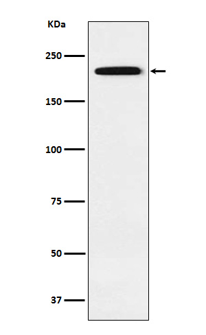
| Description | Anti-Gli3 Rabbit Monoclonal Antibody . Tested in WB, ICC/IF applications. This antibody reacts with Human. |
| Gene ID | 2737 |
|---|---|
| Other Names | Transcriptional activator GLI3, GLI3 form of 190 kDa, GLI3-190, GLI3 full-length protein, GLI3FL, Transcriptional repressor GLI3R, GLI3 C-terminally truncated form, GLI3 form of 83 kDa, GLI3-83, GLI3 |
| Calculated MW | 190 kDa |
| Application Details | WB 1:500-1:2000 ICC/IF 1:50-1:200 |
| Contents | Rabbit IgG in phosphate buffered saline, pH 7.4, 150mM NaCl, 0.02% sodium azide and 50% glycerol, 0.4-0.5mg/ml BSA. |
| Clone Names | Clone: 19G77 |
| Immunogen | A synthesized peptide derived from human Gli3 |
| Purification | Affinity-chromatography |
| Storage | Store at -20°C for one year. For short term storage and frequent use, store at 4°C for up to one month. Avoid repeated freeze-thaw cycles. |
| Name | GLI3 |
|---|---|
| Function | Has a dual function as a transcriptional activator and a repressor of the sonic hedgehog (Shh) pathway, and plays a role in limb development. The full-length GLI3 form (GLI3FL) after phosphorylation and nuclear translocation, acts as an activator (GLI3A) while GLI3R, its C-terminally truncated form, acts as a repressor. A proper balance between the GLI3 activator and the repressor GLI3R, rather than the repressor gradient itself or the activator/repressor ratio gradient, specifies limb digit number and identity. In concert with TRPS1, plays a role in regulating the size of the zone of distal chondrocytes, in restricting the zone of PTHLH expression in distal cells and in activating chondrocyte proliferation. Binds to the minimal GLI- consensus sequence 5'-GGGTGGTC-3'. |
| Cellular Location | Nucleus. Cytoplasm {ECO:0000250|UniProtKB:Q61602}. Cell projection, cilium. Note=GLI3FL is localized predominantly in the cytoplasm while GLI3R resides mainly in the nucleus (By similarity). Ciliary accumulation requires the presence of KIF7 and SMO (PubMed:19592253). Translocation to the nucleus is promoted by interaction with ZIC1 (PubMed:11238441). TMEM216 reduces its nuclear localization (By similarity). {ECO:0000250|UniProtKB:Q61602, ECO:0000269|PubMed:11238441, ECO:0000269|PubMed:19592253} |
| Tissue Location | Is expressed in a wide variety of normal adult tissues, including lung, colon, spleen, placenta, testis, and myometrium. |
Research Areas
Citations (0)

Thousands of laboratories across the world have published research that depended on the performance of antibodies from Abcepta to advance their research. Check out links to articles that cite our products in major peer-reviewed journals, organized by research category.
Submit your citation using an Abcepta antibody to
info@abcepta.com, and receive a free "I Love Antibodies" mug.
info@abcepta.com, and receive a free "I Love Antibodies" mug.

Application Protocols
Provided below are standard protocols that you may find useful for product applications.
Abcepta welcomes feedback from its customers.
If you have used an Abcepta product and would like to share how it has performed, please click on the "Submit Review" button and provide the requested information. Our staff will examine and post your review and contact you if needed.
If you have any additional inquiries please email technical services at tech@abcepta.com.
$ 185.00

⚠️ Limit: 5 vials per order for discount.
Cat# ABO15401
Ordering Information
United States
AlbaniaAustraliaAustriaBelgiumBosnia & HerzegovinaBrazilBulgariaCanadaCentral AmericaChinaCroatiaCyprusCzech RepublicDenmarkEstoniaFinlandFranceGermanyGreeceHong KongHungaryIcelandIndiaIndonesiaIrelandIsraelItalyJapanLatviaLithuaniaLuxembourgMacedoniaMalaysiaMaltaMexicoNetherlandsNew ZealandNorwayPakistanPolandPortugalRomaniaSerbiaSingaporeSlovakiaSloveniaSouth AfricaSouth KoreaSpainSwedenSwitzerlandTaiwanTurkeyUnited KingdomUnited StatesVietnamWorldwideOthers
USA Headquarters
(888) 735-7227 / (858) 622-0099 or (858) 875-1900
Other Products
Shipping Information
Domestic orders (in stock items)
Shipped out the same day. Orders placed after 1 PM (PST) will ship out the next business day.
International orders
Contact your local distributors
